


Review
History
Medication: Captopril, Amlodipine, Atorvastatin.
PE: (-)
药物:卡托普利,氨氯地平,阿托伐他汀。
 Figure 1. MRA revealed a left A2/3 bifurcation aneurysm with an irregular shape.
Figure 2. DSA showed the right ACA A1 segment was undeveloped and the right ICA clinoid segment mild/moderate stenosis.
图 2. 脑血管造影示右侧大脑前动脉A1段发育不佳,右侧颈内动脉床突段轻中度狭窄。
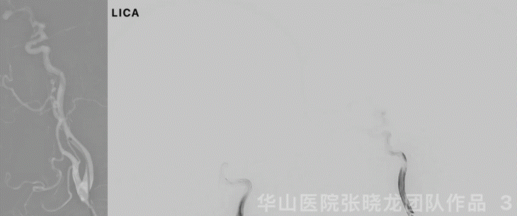
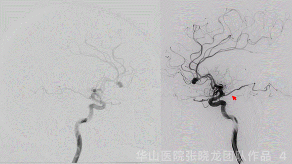
Figure 4 GIF. Rotational DSA confirmed a left A2/3 irregular aneurysm, a small anterior choroidal artery aneurysm and an ophthalmic aneurysm. The thick artery (arrow) was the left anterior choroidal artery instead of posterior communicating artery.
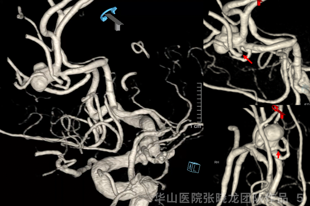
Figure 5 GIF. 3D reconstructions showed the three aneurysms all irregular and a branch originating from the A2/3 aneurysm neck, which should be preserved intra-operatively.
1
Strategy
ØIndications and strategies
1、For the A2/3 bifurcation dissecting aneurysm :
•Considering the elderly and the acute curve of the A2/3, simple coiling is preferable to stent-assisted coiling, which can cause thrombus or occlusion of the left ACA.
•The left ACA occlusion would be a disaster due to the ipsilateral MCA superior trunk occlusion.
•A branch originates from the aneurysm neck, which should be preserved intra-operatively.
b.A single large coil technique is preferred to preserve the branch or decrease the recurrence rate.
2、For the irregular small anterior choroidal artery aneurysm:
•The aneurysm neck initiates from the anterior choroidal artery that should be preserved during coiling.
3、For the relatively irregular ophthalmic aneurysm:
Ø指征和策略
1、A2/3分叉部夹层动脉瘤治疗策略:
•患者高龄,A2/3段成角较急,支架辅助栓塞大脑前动脉血栓形成或闭塞风险相对较高,故首选单纯栓塞。
•由于同侧大脑中动脉上干闭塞,主要由同侧大脑前动脉软膜吻合代偿,所以若术中导致左侧大脑前动脉闭塞将会导致灾难性的后果。
•瘤颈部有一支血管发出,术中栓塞时应注意保护。保护方式如下:
•动脉瘤瘤颈部不必致密栓塞。
•采用单圈大圈技术,保护血管的同时降低复发风险。
2、不规则脉络膜前动脉小动脉瘤治疗策略:
•脉络膜前动脉发自动脉瘤瘤颈部,栓塞时应保护粗大的脉络膜前动脉。
3、不规则颈眼动脉瘤治疗策略:
•宽颈颈眼动脉瘤采用支架辅助栓塞。
2
Strategy
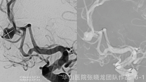

图 6 GIF. 测量动脉瘤大小7.9*7.8mm,瘤颈3.7mm。行全身肝素化。由于髂动脉和腹主动脉迂曲,选用90cm 0.089长鞘置于左侧颈内动脉起始部。115cm 6F通桥中间导管置于颈内动脉颅底处。经导管灌注尼莫地平1ml。直头SL-10微导管在微导丝导引下置于瘤腔。经微导管依次填入4枚弹簧圈(Tonbridge Feng helical 10mm*30cm, 7mm*30cm, 6mm*20cm & Target 360 3mm*4cm) 。
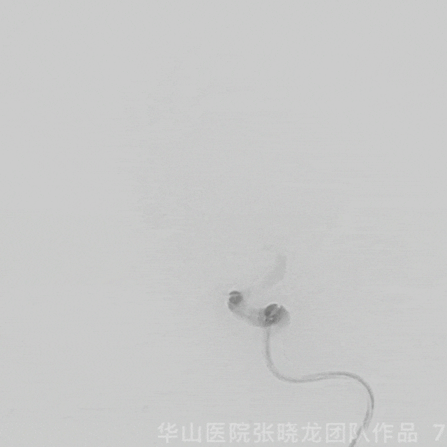
Figure 7 GIF. Considering the pericallosum artery and the perforators from aneurysm neck, plus the patient’s age, though DSA showed the A2/3 aneurysm residual, the operation was finished. And the intracranial vessels were patent.
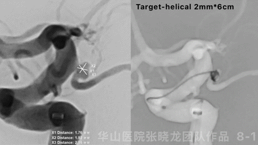

Figure 9. Measurements: An size 2.6*2.1mm, neck 2.1mm, proximal parent artery diameter 4.05mm, distal parent artery diameter 3.95mm. XT-27 was placed into the left ACA, a spiral-curved Echelon-10 was placed into the sac. Deployed a Neuroform EZ 4*15mm. Then two coils (Target-360 2mm*3cm/1.5mm*3cm) was inserted.

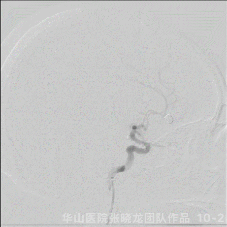
Figure 10 GIF. Angiograms showed the aneurysms were packed satisfactorily while the left anterior choroidal artery was not revealed.
Figure 11 GIF. Dyna-CT did not demonstrate any bleeding. Tirofiban 6ml and Nimodipine 1ml were administered.

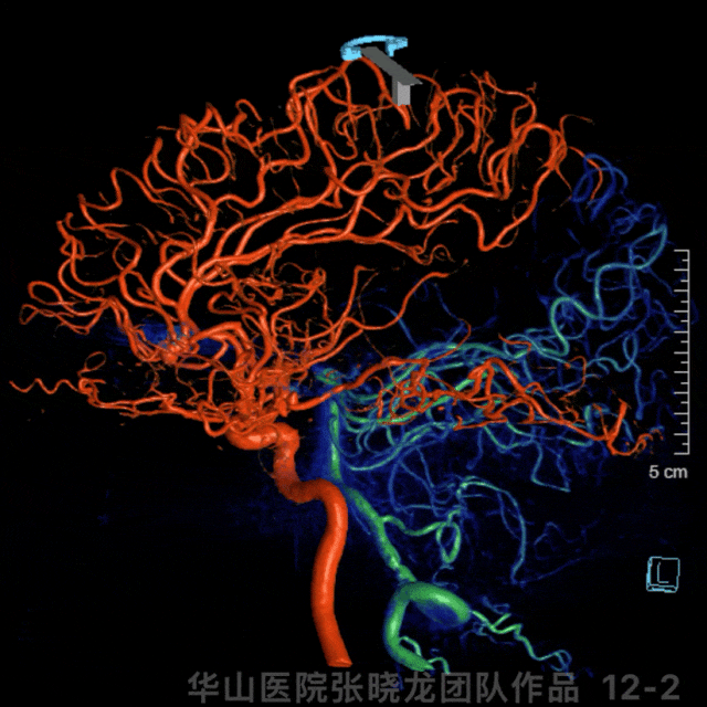
Figure 14 GIF. Dyna-CT did not detect hemorrhage.
3
Post-Operation
•Medication:
1、Tirofiban 10ml/h was prescribed for 36 hours.
2、Aspirin 100mg qd and Clopidogrel 75mg qd were prescribed after the operation.
3、Clopidogrel Gene was normal.
4、ADP 96.1%, AA 100%.
•药物:
1、替罗非班10ml/h维持36h。
2、术后予阿司匹林100mg qd和氯吡格雷75mg qd口服。
3、氯吡格雷基因代谢正常。
Video 1. Minor ischemic high signal of the left temporal lobe. The anterior choroidal artery is patent from the MRA.
视频 1. 术后复查头颅磁共振,DWI示左侧颞叶少许急性脑梗灶,MRA提示脉络膜前动脉通畅。
4
Summary
ØIndications and strategies
1、For the A2/3 bifurcation dissecting aneurysm :
•Considering the elderly and the acute curve of the A2/3, simple coiling is preferable to stent-assisted coiling, which can cause thrombus or occlusion of the left ACA.
•The left ACA occlusion would be a disaster due to the ipsilateral MCA superior trunk occlusion.
•A branch originates from the aneurysm neck, which should be preserved intra-operatively.
b.A single large coil technique is preferred to preserve the branch or decrease the recurrence rate.
2、 For the irregular small anterior choroidal artery aneurysm:
•The aneurysm neck initiates from the anterior choroidal artery that should be preserved during coiling.
3、 For the relatively irregular ophthalmic aneurysm:
Ø指征和策略
1、 A2/3分叉部夹层动脉瘤治疗策略:
•患者高龄,A2/3段成角较急,支架辅助栓塞大脑前动脉血栓形成或闭塞风险相对较高,故首选单纯栓塞。
•由于同侧大脑中动脉上干闭塞,主要由同侧大脑前动脉软膜吻合代偿,所以若术中导致左侧大脑前动脉闭塞将会导致灾难性的后果。
•瘤颈部有一支血管发出,术中栓塞时应注意保护。保护方式如下:
•动脉瘤瘤颈部不必致密栓塞。
•采用单圈大圈技术,保护血管的同时降低复发风险。
2、不规则脉络膜前动脉小动脉瘤治疗策略:
•脉络膜前动脉发自动脉瘤瘤颈部,栓塞时应保护粗大的脉络膜前动脉。
3、不规则颈眼动脉瘤治疗策略:
声明:脑医汇旗下神外资讯、神介资讯、脑医咨询、Ai Brain 所发表内容之知识产权为脑医汇及主办方、原作者等相关权利人所有。
投稿邮箱:NAOYIHUI@163.com
未经许可,禁止进行转载、摘编、复制、裁切、录制等。经许可授权使用,亦须注明来源。欢迎转发、分享。
投稿/会议发布,请联系400-888-2526转3。

