Review
History
mild cognitive disorder: MMSE 25/30, MoCA 23/30 THI 0/100
• Pre-operative medication: Rivaroxaban 10mg QD; ticagrelor 90mg BID.
认知功能轻度下降:MoCA 23/30,MMSE 25/30。 耳鸣致残量表 0/100。


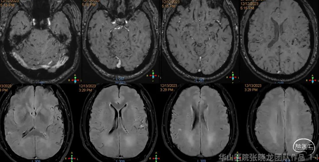
Figure 1. MRA revealed increased dilated vessels at the bilateral lateral sinuses. SWI did not detect any obvious micro-bleedings.
图 1. MRA提示双侧侧窦区增多增粗血管影。SWI未见明显微出血灶。

Figure 2 GIF. Right MMA and occipital feeders converged to the right transverse-sigmoid junction (Cognard I), while the sinus septum could be identified on the proximal side. Right TS was not revealed via RECA. The MMA was regraded as “golden access” for trans-arterial embolization.


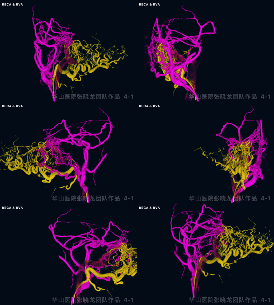
Figure 4 GIF. RECA fusion 3D reconstruction, revealed fistulae concentrated to the transverse-sigmoid junction. (internal maxillary artery (purple) and occipital artery (yellow); internal maxillary artery (white) and right vertebrate artery (green)).

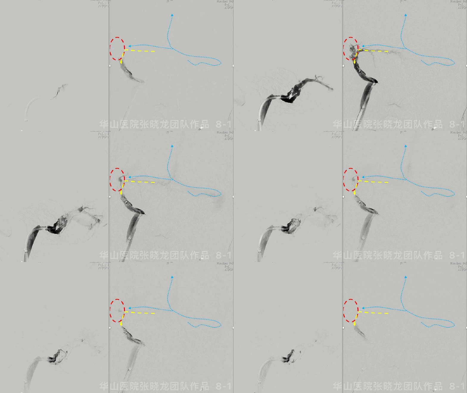
Figure 5 GIF. Fistula located at left transverse-sigmoid junction, emptied to the TS, SSS, SS, right TS and cortical veins (Cognard 2a+b). The LEFT sigmoid sinus occluded, while the right transverse-sigmoid junction also obstructed (red lines).
图 5 GIF. 左侧颈外动脉分支向左侧横-乙交界区的汇聚,再通过左侧横窦向窦汇、上矢状窦、直窦以及右侧横窦残端引流(Cognard 2a+b) 左侧乙状窦闭塞(红虚线)位于DAVF下游;右侧横-乙交界区闭塞(位于DAVF上游)。


1
Bilateral DAVFs angioarchitecture analysis
Angioarchitecture of the right lateral sinus DAVF
1. Cognard I, sinus type;
4. Right transvers-sigmoid junction sinus occlusion.
Angioarchitecture of left transverse-sigmoid junction DAVF
1. Cognard IIa+b, sinus type;
4. Retrograde drainage of pial veins interfered the judgement of focal angioarchitecture.
右侧侧窦区DAVF血管构筑
1. Cognard I,窦型;
4. 右侧横-乙状窦交界区静脉窦闭塞。
左侧横-乙交界区DAVF的血管构筑
1. Cognard IIa+b,窦型;
4. 经横窦向软膜静脉窦逆流,静脉结构迂曲扩张,干扰对局部动静脉瘘血管构筑的判断。


2
First stage strategies
Flow reduction and venous sinus reconstruction
1. The main target for the bilateral transverse-sigmoid junction DAVFs with bilateral occluded sinuses was sinus recanalization in order to reconstruct anterograde drainage instead of simple fistula obliteration.
2. Right transverse-sigmoid sinus recanalization:
The occluded segment was located in the upstream of the fistula, which was a protective factor for right DAVF while aggravated left side drainage disorder. Therefore the occluded sinus should be recanalized.
Consider the short obstructed segment on the right side, the recanalization might be technically easier.
Antegrade recanalization from right to the left side was technically preferable.
Transvenous access route can obliterate the shunted pouch effectively.
3. Left sigmoid sinus recanalization:
Consider the dominant size of the left sinus, sinus reconstruction was prior to sacrifice.
Left jugular foramen was involved, therefore retrograde recanalization through left internal jugular vein was technically difficult.
The long-term risk of jugular foramen intra-stent stenosis was high.
If the left sinus reconstruction failed, the left sinus sacrificed and right sinus reconstruction can provide enough venous drainage capacity.
1. 双侧横-乙交界区DAVF伴有双侧静脉窦闭塞,重建静脉窦的正向回流是主要目标,而非单纯闭塞动静脉瘘;
2. 右侧横-乙交界区闭塞再通:
静脉窦闭塞段位于动静脉瘘汇聚区的上游,是相对右侧DAVF的保护性因素,但导致左侧DAVF的静脉回流障碍进一步加重,仍有干预的指征;
右侧静脉入路相对更稳定,横-乙交界静脉窦闭塞节段短,再通重建的技术难度相对于左侧低,静脉窦远期通畅率高;
右侧入路正向开通左侧闭塞乙状窦技术成功率更高;
右侧静脉入路可能直接进入动静脉瘘硬膜血管汇集区,可以经静脉途径高效地靶向栓塞DAVF。
3. 左侧乙状窦闭塞再通:
左侧侧窦优势侧,仍倾向于静脉窦重建优于闭塞静脉窦;
左侧闭塞段累及乙状窦颈静脉孔段,经左侧颈内静脉逆行开通技术难度大;
左侧乙状窦经静脉孔段支架远期再狭窄风险;
双侧静脉窦至少需要确保重建一侧的正向回流,当左侧静脉窦再通失败时,经静脉途径闭塞静脉窦也是备用方案。


Red circle on the lateral side was fed by the ECA feeders and trans-arterial embolization through MMA was technically not too hard;
Yellow dot line indicated the PMA fed torcular fistula. Large balloon to protect sinus was required if MMA was selected, which will increase the risk of normal drainage vein obliteration and onyx refluxing to arteries.
The 3D roadmap can separate the right TS (blue dot lines)from the fistulous veins.
外侧静脉腔隙(红圈)由颈外动脉分支主要参与供血, 可以通过脑膜中动脉入路闭塞,技术难度最低; 内侧静脉腔隙(黄色虚线)通过脑膜后动脉供血。如果经通过脑膜中动脉,则需要静脉窦大球囊保护,增加Onyx胶沿球囊闭塞功能引流静脉或向动脉端危险返流的风险。
通过双侧颈外动脉三维容积成像的融合,可以显示上游的横窦的走行(蓝色虚线)。


Best overall perspective of venous structure;
Three retrograde transvenous routes:
1. dural veins from PMA (green)
2. veins from MMA (lateral)
3. occluded sinus (blue)
Torcular fistula would be difficult to manage via bilateral MMA routes. Therefore TVE of the torcular fistula is preferred.
动-静脉联合路途显示整体血管构筑; 经静脉逆行有三个路径:
1. 脑膜后动脉来源的硬膜静脉(绿)
2. 脑膜中动脉来源(外侧)
3. 闭塞的静脉窦(蓝);
脑膜后动脉的供血来源(绿色)经动脉栓塞相对困难,而经静脉可以靶向闭塞静脉汇聚区。
3
Sequence of TVE, TAE and sinus recanalization:
2. MMA is the golden route for second staged trans-arterial embolization.
3. Right occluded sinus recanalized first to reconstruct left/bilateral cerebral drainage veins.
2. MMA是二期经动脉治愈性栓塞的最优通路;


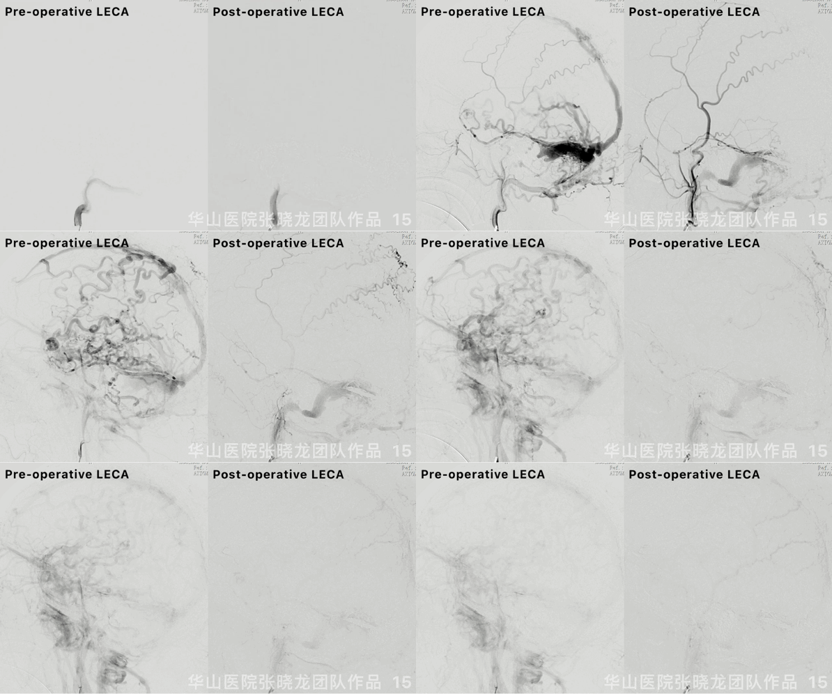
图 13 GIF. 左侧静脉窦重建后,Cognard IIa+b DAVF转变为低分级Cognard I级。

图 14. Precise 7*30mm和7*40mm支架于右侧横乙交界区释放。LitePac 6*30mm球囊以6-8ATM扩张支架使支架贴壁良好。

4
Post-operation
• Medication: aspirin and clopidogrel.
• 术前 MoCA 23/30, 术后 MoCA 26/30
• 药物:口服阿司匹林和氯吡格雷。
声明:脑医汇旗下神外资讯、神介资讯、脑医咨询、Ai Brain 所发表内容之知识产权为脑医汇及主办方、原作者等相关权利人所有。
投稿邮箱:NAOYIHUI@163.com
未经许可,禁止进行转载、摘编、复制、裁切、录制等。经许可授权使用,亦须注明来源。欢迎转发、分享。
投稿/会议发布,请联系400-888-2526转3。


