Review
History
• 63 y/o male.
• Suffered from dizziness, unsteady gait and double vision for 3 months.
• Past medical history: HTN for 30 years; smoking for 30 years, quitted for 3 months; drinking for 30 years, not quitted.
• Medication: Aspirin, Clopidogrel, Atorvastatin and Valsartan.
• NE: (-)
• 63岁,男性。
• 头晕、行走不稳伴视物模糊3月。
• 既往史:高血压30年;吸烟30年,已戒3月;饮酒30年,未戒酒。
• 药物:阿司匹林,氯吡格雷,阿托伐他汀和缬沙坦。
• 神经查体:-。


Figure 1 GIF. TOF MRA revealed right ICA cavernous segment stenosis and MRI revealed pons and cerebral peduncle infarctions.
图 1 GIF. TOF MRA提示右侧颈内动脉海绵窦段狭窄,MRI提示脑桥脑和大脑脚梗死。

Figure 2 GIF. No microbleedings were observed from SWI.
图 2 GIF. SWI未见明显微出血。

Figure 3. CTP revealed right cerebral hemisphere hypoperfusion.
图 3. CTP提示右侧大脑半球低灌注。

Figure 4. Severe stenosis was found at right undominant vertebral artery initial segment and the terminal segment occluded.
图 4. DSA造影证实右侧劣势椎动脉起始段重度狭窄,V4段以远闭塞。

Figure 5. Left dominant vertebral artery V4 segment almost occluded and basilar artery was supplied by anastomoses.
图 5. 左侧优势椎动脉V4段夹层伴重度狭窄近闭塞,远端通过右侧小脑后下动脉及软膜血管代偿。
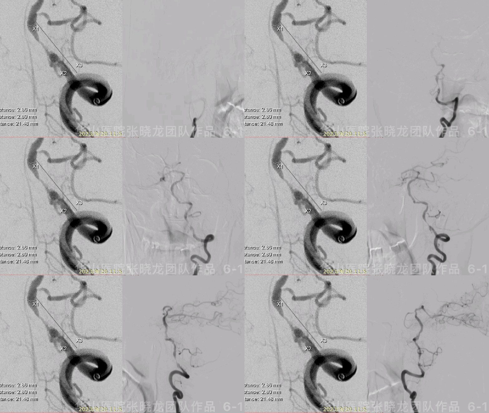

Figure 6 GIF. Dissecting and severe stenosis of left V4 segment was observed and left PCOM arose nearby.
图 6 GIF. 旋转造影及3D重建示左椎V4段夹层伴线样狭窄,邻近发出左侧小脑后下动脉。

Figure 7. Tortuous left ICA compensated posterior circulation and right anterior circulation.
图 7. 左侧颈内动脉迂曲,代偿后循环和右侧大脑半球。
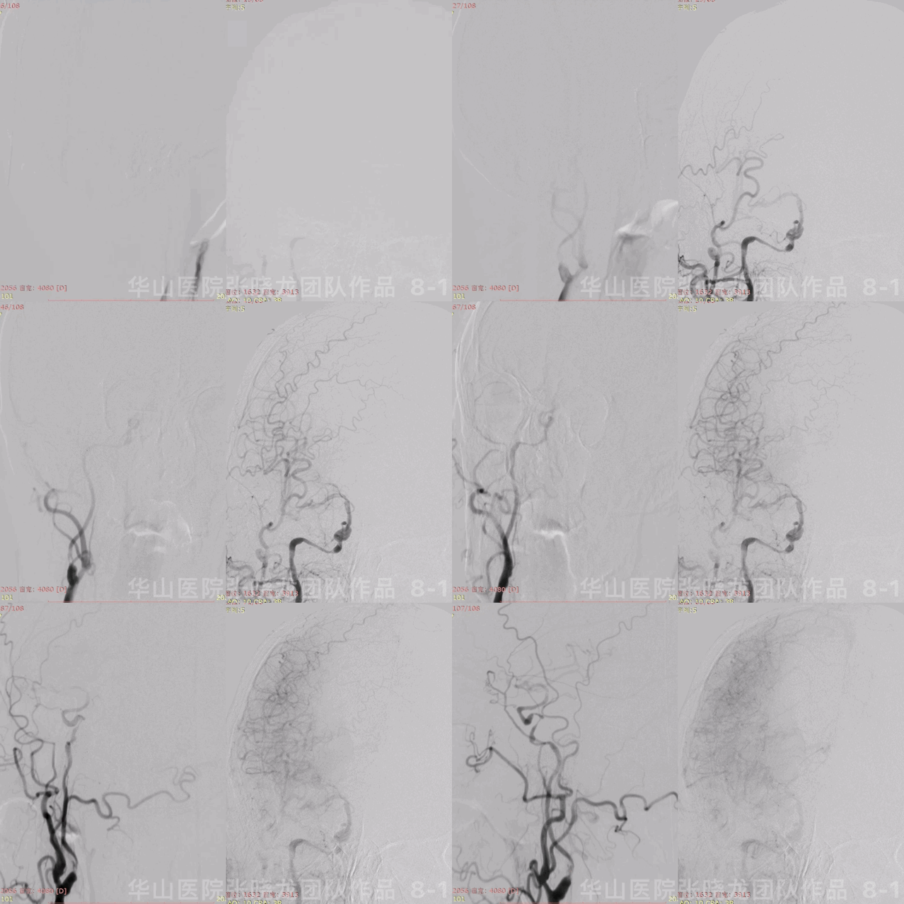

Figure 8 GIF. DSA depicted right ICA initial segment and cavernous segment severe stenosis and the artery extremely serpentine.
图 8 GIF. 造影示右侧颈内动脉起始部及海绵窦段重度狭窄,颈内动脉极其迂曲。
1
Strategy
1.Chronic infarctions were found in the cerebellar and pons.
2.Undominant right vertebrate artery terminal segment was occluded and initial segment severe stenosis.
3.Dominant left vertebrate artery V4 segment occluded and anastomoses compensated the VA. Bilateral ICA can not supply basilar artery, therefore dominant left vertebrate artery V4 segment should be recanalized.
4.Right ICA initial segment almost occluded that should be recanalized first, while the severe stenosis was dilated by balloon only to reduce hyper-perfusion risk. The cavernous segment can be performed angioplasty at the second stage.
5.Systolic blood pressure was monitored between 100-120mmHg.
1.磁共振提示大脑脚及脑桥陈旧性脑梗死。
2.右侧劣势椎动脉远端闭塞,起始部重度狭窄。
3.左侧优势椎动脉v4段闭塞,通过吻合支代偿。基底动脉主要通过左侧小脑后下动脉显影,双侧颈内动脉无明显代偿。所以左侧优势椎动脉V4段建议开通。
4.右侧颈内动脉起始部和海绵窦段重度狭窄近闭塞,一期先开通右侧颈内动脉起始部,二期再行海绵窦段血管成形术。为减少术后高灌,起始部重度狭窄仅行球囊扩张。
5.术后收缩压控制在100-120mmhg。
2
Operation


Figure 9 GIF. General heparinization was performed. 80cm 6F NeuroMax was placed at left VA initial segment with the support of 125cm MP and 0.035 microwire. Nimodipine 1ml was administered. 6F 105cm Tonbridge catheter was positioned at left V4 segment. Echelon-10 with a straight tip was advanced through the stenosis via Synchro-II microwire. Angiograms confirmed the microwire in the real lumen. Floppy-300 microwire was navigated into basilar artery and then dilated Gateway 2.75*15mm balloon 6ATM for 10s. Angiograms showed the stenosis was expanded satisfactorily without dissecting formation. Withdrew the balloon and microwire. Deployed a Wingspan 3.0*20mm.
图 9 GIF. 行全身肝素化。将80cm 6F NeuroMax长鞘在125cm MP和0.035导丝支撑下置于左椎起始部,灌注尼莫地平1ml。6F 105cm Tonbridge中间导管置于左椎V4段。直头Echelon-10微导管在Synchro-II微导丝支撑下通过狭窄段。手推造影证实微导管位于真腔内。更换Floppy-300微导丝置于基底动脉近端,Gateway 2.75*15mm球囊于狭窄段6ATM扩张10s,复查造影狭窄段扩张满意,未见明显出血或新发夹层形成。撤回球囊及微导丝。于狭窄段释放Wingspan 3.0*20mm支架。
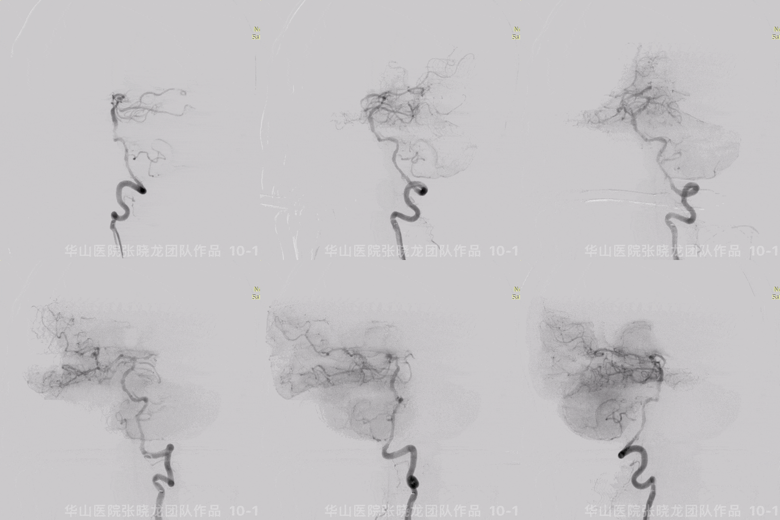
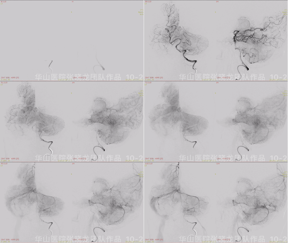
Figure 10 GIF. Left V4 segment stenosis was dilated satisfactorily and posterior circulation blood flow improved. Tirofiban 6ml and Nimodipine 0.5ml were administered.
图 10 GIF. 复查造影左椎V4段狭窄扩张满意,后循环灌注增加。经导引导管予替罗非班6ml和尼莫地平0.5ml。

Figure 11. 80cm 6F NeuroMax was placed at right common carotid artery with the support of 125cm MP and 0.035 microwire. 100cm MP was advanced through the stenosis via Synchro-II microwire. Angiograms confirmed the microcatheter in the real lumen. Floppy-300 microwire was navigated into right cavernous segment and deployed Spider 5mm at the cavernous segment. Dilated Gateway 3.5*15mm balloon from far to near.
图 11. 将80cm 6F NeuroMax长鞘在125cm MP和0.035导丝支撑下置于右侧颈总动脉。100cm MP微导管在Synchro-II微导丝支撑下通过狭窄段,路途证实微导管位于真腔内。更换Floppy-300微导丝置于右侧颈内动脉海绵窦段,Spider 5mm保护伞在海绵窦段释放。Gateway 3.5*15mm球囊于狭窄段由远及近扩张。

Figure 12 GIF. The initial segment severe stenosis dilated satisfactorily though remained moderate stenosis.
图 12 GIF. 复查造影右侧颈内动脉起始部重度狭窄扩张满意,残留中度狭窄。
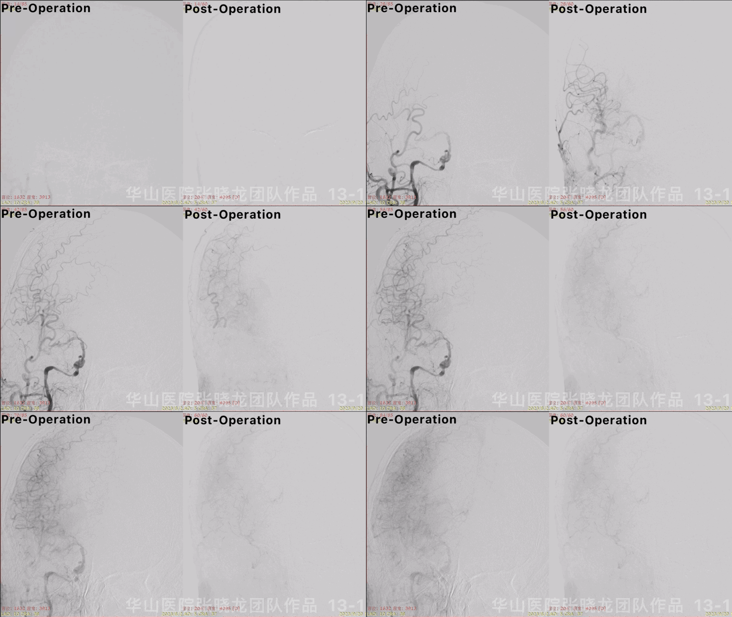

Figure 13 GIF. Angiograms showed blood flow increased and intracranial vessels were patent.
图 13 GIF. 复查造影右侧大脑半球血流改善,未见颅内血管出血或栓塞。

Figure 14 GIF. Dyna-CT did not reveal any hemorrhage.
图 14 GIF. 术后即刻Dyna-CT未见出血。
3
Post-Operation
NE:
GCS 15, normal pupils movement and light reflux, normal muscle strength, no swallow difficulty, no new neurologic defects.
Medication:
Nimodipine was administered, systolic blood pressure was controlled between 100mmHg and 140mmHg.
Continue Aspirin and Clopidogrel. (AA 92.3%, ADP 60.9%)
查体:
GCS 15,双侧瞳孔等大等圆,对光反射灵敏,眼球各项运动正常,四肢肌力正常,无吞咽困难,无新发神经功能缺损。
药物:
予尼莫地平,收缩压控制在100mmHg and 140mmHg。
继续阿司匹林及氯吡格雷。(阿司匹林抑制率92.3%,氯吡格雷抑制率60.9%。

Figure 15. DWI revealed acute dotted infarctions on the right paraventricular.
图 15. 术后第一天复查磁共振提示右侧侧脑室旁点状急性脑梗死。

Figure 16. CTP improved after operation.
图 16. 术后第7天复查头颅灌注脑灌注改善,双侧大脑半球灌注基本对称。

Figure 17. MRI depicted hemorrhage one the right temporal lobe. DWI did not detect any new infarctions. No new neurologic defects occurred. Stopped Clopidogrel and continue Aspirin. Controlled systolic blood pressure between 100-120mmHg.
Figure 17. 术后17天复查MRI提示右侧颞叶出血,DWI未见新发脑梗死。患者无新发神经功能缺损症状。暂停氯吡格雷,继续口服阿司匹林,控制收缩压100-120mmhg。
4
Summary
1.Chronic infarctions were found in the cerebellar and pons.
2.Undominant right vertebrate artery terminal segment was occluded and initial segment severe stenosis. Right vertebrate artery supplied right posterior inferior cerebellar artery. The stenosis was recommended follow up. If the stenosis became more severe, angioplasty can be performed.
3.Dominant left vertebrate artery V4 segment occluded and anastomoses compensated the VA. Microcatheter can not be navigated into the distal vessels through anastomoses which may lead to hemorrhage during the procedure. Bilateral ICA can not supply basilar artery, therefore dominant left vertebrate artery V4 segment should be recanalized.
4.Right ICA initial segment almost occluded that should be recanalized first, while the severe stenosis was dilated by balloon. The cavernous segment can be performed angioplasty at the second stage. Meanwhile post-operative systolic blood pressure was strictly controlled. All above measures intended to reduce post-operative hyper-perfusion.
5.Although staged treatments were adopted, hyper-perfusion still occurred. Luckily, no severe complications happened. Therefore, staged treatments were significant especially for severe stenosis.
1.磁共振提示大脑脚及脑桥陈旧性脑梗死。
2.右侧劣势椎动脉远端闭塞、起始部重度狭窄,右侧椎动脉发出右侧小脑后下动脉,右椎动脉可随访。若狭窄进一步加重,建议行血管成形术。
3.左侧优势椎动脉v4段闭塞,通过吻合支代偿,术中操作时,微导管不能走吻合血管到达远端,否则扩张会导致吻合血管破裂。基底动脉主要通过左侧小脑后下动脉显影,双侧颈内动脉无明显代偿。所以左侧优势椎动脉V4段建议开通。
4.右侧颈内动脉起始部和海绵窦段重度狭窄近闭塞,一期先行右侧颈内动脉起始部单纯球囊扩张术改善颅内血流,二期再行海绵窦段血管成形术。同时术后严格控制收缩压。这些策略都是为了降低术后高灌注风险。
5.即便我们已经采取了分期治疗,还是发生了术后高灌注的出血,然而并没有产生致死性的后果,这说明了对于严重狭窄病例的分期治疗十分必要。
张晓龙
复旦大学附属华山医院
复旦大学附属华山医院放射科主任医师,博士、教授、博士生导师;
斯坦福大学医学院客座临床教授;
主持国家自然科学基金3项,第一作者或通讯作者发表国内外权威期刊文章50余篇;
中华医学会、放射学会、卫生部医政司等组织中担任副主任委员、组长等职务.《中国名医百强榜》神经介入专业中国十强(2012年度、2013年度、2014年度、2015-16年度、2017-18年度);
擅长复杂和疑难脑血管疾病的介入治疗,如复杂脑动脉瘤的栓塞,硬脑膜动静脉瘘栓塞,脑动静脉畸形栓塞,脑梗死的支架,脊髓血管畸形治疗;
自1995年开始从事脑血管疾病介入诊治工作和研究,师从黄祥龙教授、沈天真教授和凌锋教授,是我国最早从事神经介入的专家之一。2010年9月至今连续介入治疗颅内动脉瘤1500余例,无操作致死.
点击上方二维码
查看更多“介入”内容
声明:脑医汇旗下神外资讯、神介资讯、脑医咨询、Ai Brain 所发表内容之知识产权为脑医汇及主办方、原作者等相关权利人所有。
投稿邮箱:NAOYIHUI@163.com
未经许可,禁止进行转载、摘编、复制、裁切、录制等。经许可授权使用,亦须注明来源。欢迎转发、分享。
投稿/会议发布,请联系400-888-2526转3。


