Review
History
• 59 y/o female.
• Local hospital CTA revealed a right posterior communicating artery aneurysm accidentally.
• Past medical history: DM, No HTN, No smoking cigarettes and/or alcohol consumption.
• Medication: Atorvastatin, Epperidone, Mecobalamin and Repaglinide.
• NE:(-).
• 59岁,女性。
• 药物:阿托伐他汀,乙哌立松,甲钴胺,瑞格列奈。
• 神经查体:-。

图 1. DWI未见急性脑梗灶。


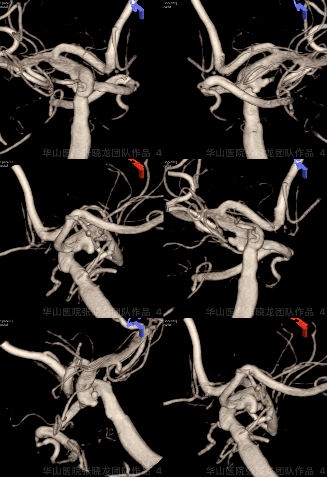
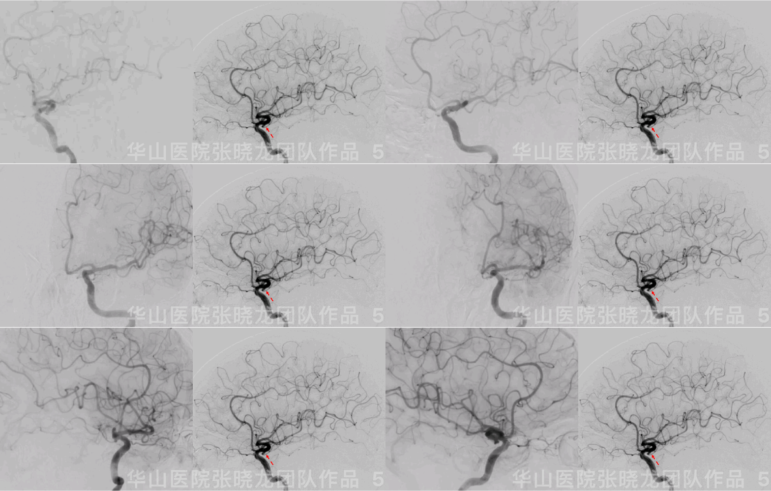
Figure 3 GIF. DSA confirmed a right fetal-type posterior communicating artery aneurysm with a relative wide neck. Right P1 segment was undeveloped.
图 3 GIF. DSA证实右侧胚胎型后交通宽颈动脉瘤。右侧P1段未发育。
1
Strategy
A fetal-type posterior communicating artery aneurysm with multiple daughter sacs should be treated.
Right P1 segment were undeveloped. Therefore the right Pcom artery must be preserved.
Multiple daughter sacs should be recognized in working projectings to avoid intraoperative rupture and protect fetal-type posterior communicating artery meanwhile.
Atlas stent can be selected for protecting posterior communicating artery via Swinging-Tail technique.
T-stent technique was scheduled:
XT-27 stenting microcatheter was placed in ICA; SL-10 stenting microcatheter in posterior communicating artery placed far for Atlas deployment.
Neuroform stent was deployed for ICA.
胚胎型后交通动脉瘤伴子瘤破裂风险高,建议治疗。
原始后交通发自动脉瘤颈,右侧P1段不发育,因此右侧后交通动脉(胚胎型)必须保护。
选择合适的工作角度(多个工作角度),能清楚显示子瘤和动脉瘤颈部。
为保护后交通动脉,可采用Atlas “神龙摆尾” 技术。
计划采用T型支架技术:
XT-27支架微导管置于颈内动脉,SL-10微导管置于后交通动脉内用于释放Atlas支架。
颈内动脉内释放Neuroform支架。
2
Operation

Figure 6. Measurements: size 2.46*3.33mm, neck 2.21mm, proximal parent artery diameter 3.4mm, distal parent artery diameter 3.3mm. 6F Envoy DA guiding catheter was placed into the right cavernous segment. SL-21 microcatheter was advanced into the right posterior communicating artery. Then changed to another working projection. XT-27 was placed into the right M1 segment and deployed Neuroform 3.5*15mm stent.
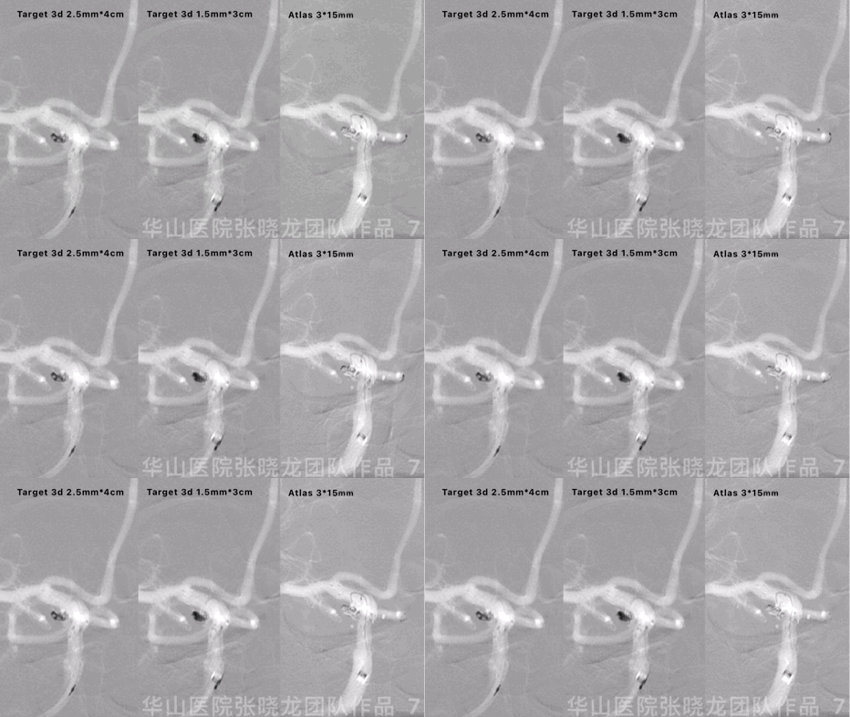
Figure 7 GIF. C-curved Echelon-10 was navigated into the sac to inserted Target 3d 2.5mm*4cm and Target 3d 1.5mm*3cm coils. General heparization was conducted. Then an Atlas 3*15mm was deployed into the right posterior communicating artery.
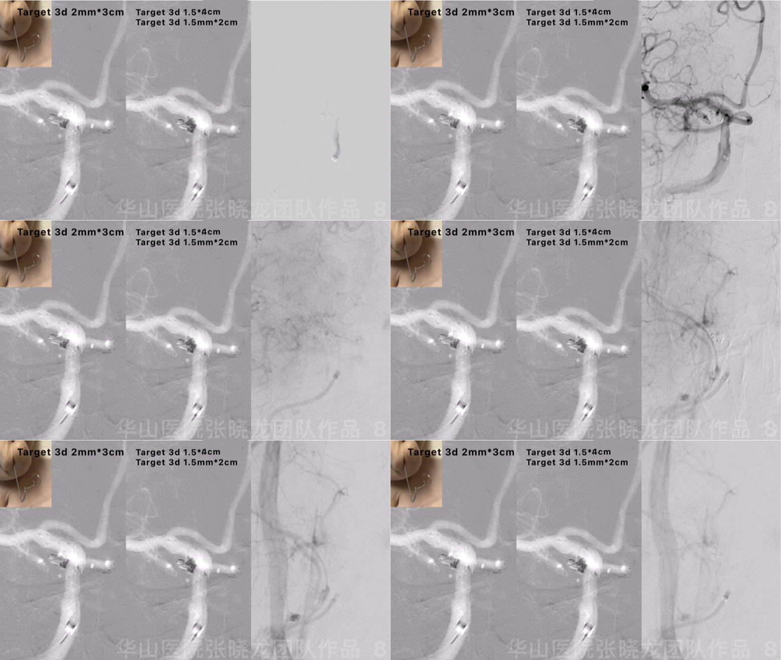
Figure 8 GIF. Lateral spiral curved Echelon-10 was re-navigated into the sac and inserted 3 coils (Target 3d 2mm*3cm, 1.5mm*4cm, 1.5mm*2cm). Angiograms showed the aneurysm was densely packed and parent artery was patent.
图 8 GIF. Echelon-10重新塑螺旋形后超选入动脉瘤腔内,依次填入3枚弹簧圈(Target 3d 2mm*3cm, 1.5mm*4cm, 1.5mm*2cm)。复查造影动脉瘤致密栓塞,载瘤动脉通畅。
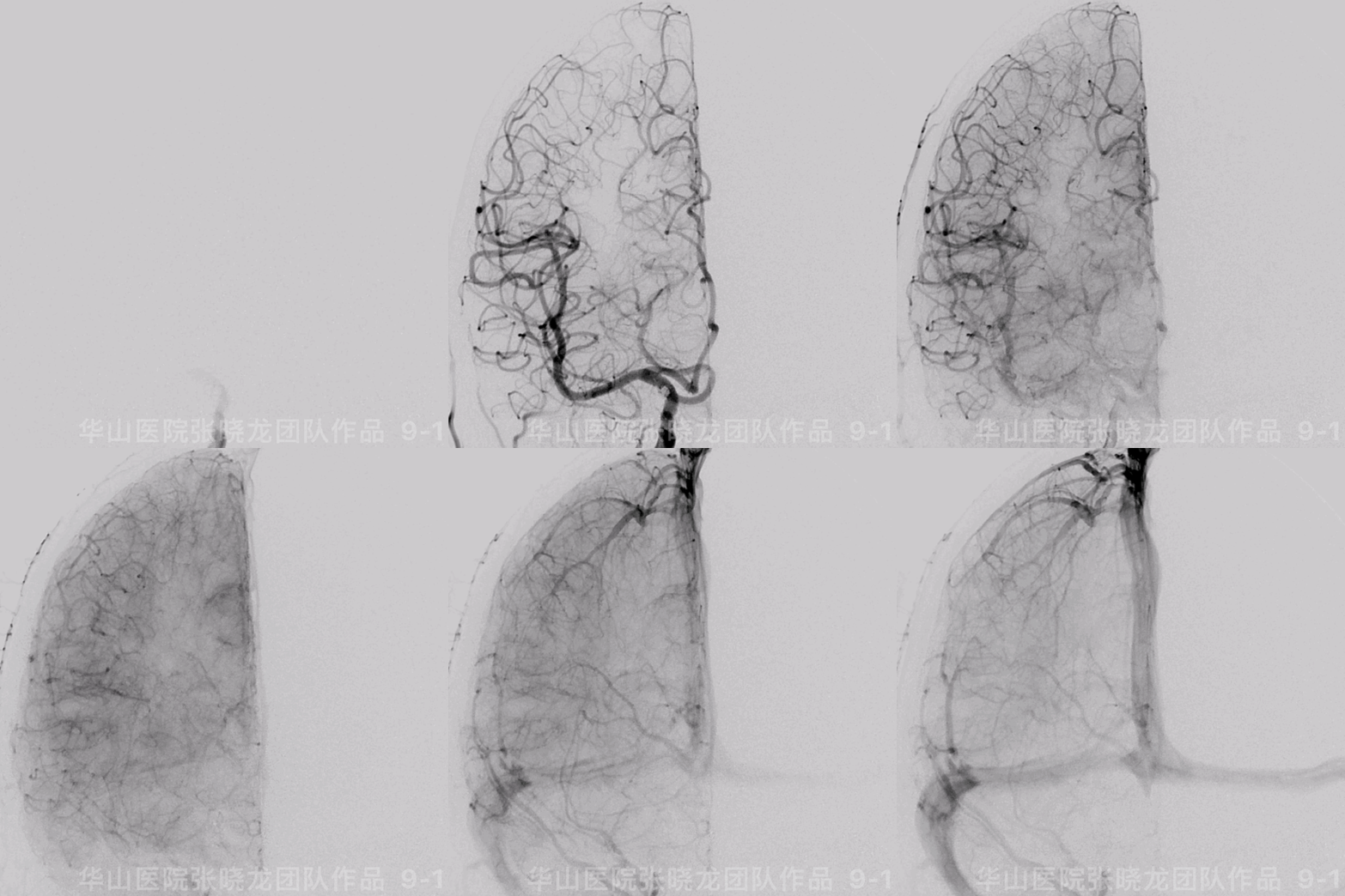
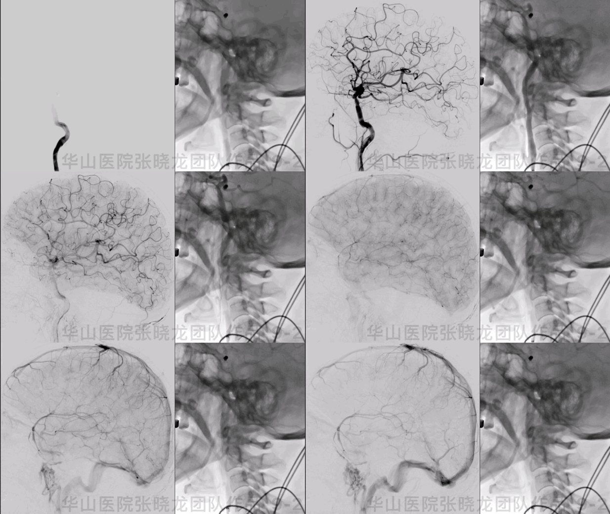
Figure 9 GIF. The aneurysm was packed satisfactorily and the intracranial vessels were intact.
图 9 GIF. 复查造影示动脉瘤栓塞满意,颅内血管完好。

Figure 10 GIF. Dyna-CT did not demonstrate hemorrhage. No acute infarctions were found from post-operative day 4 DWI.
3
Post-Operation
Medication: Tirofiban maintained for 48 hours.
At discharge: Aspirin for long-term and Ticagrelor for 3 months.
神经查体:
GCS 15,双侧眼球运动正常,对光反射灵敏,言语清,四肢肌力正常,双侧巴氏症阴性。
出院:阿司匹林长期口服,替格瑞洛口服3月。
4
Summary
A fetal-type posterior communicating artery aneurysm with multiple daughter sacs should be treated.
Due to minor aneurysm with multiple sac, large coils to apposition was difficult to form. Small 3D coils were selected (while small coils were easily compressed).
Heparin timing was scheduled after farming coil placed.
Right P1 segment were undeveloped. Therefore the right Pcom artery must be preserved.
Atlas stent can be selected for protecting posterior communicating artery via Swinging-Tail technique. Solitaire stent was an alternative to protect Pcom artery.
T-stent technique was scheduled:
XT-27 stenting microcatheter was placed in ICA; SL-10 stenting microcatheter in posterior communicating artery placed far for Atlas deployment.
Neuroform stent was deployed for ICA.
Coiling aneurysm through meshing technique.
胚胎型后交通动脉瘤伴子瘤破裂风险高,建议治疗。
后交通动脉瘤较小且多发子瘤,大圈成篮困难,小的3d的圈可以更好的栓塞(但小圈容易压缩复发)。
肝素化时机非常重要,选择在弹簧圈成篮后行肝素化。
原始后交通发自动脉瘤颈,右侧P1段不发育,因此右侧后交通动脉(胚胎型)必须保护。
为保护后交通动脉,Atlas支架释放时可采用“神龙摆尾”技术。当然也可以选择Solitaire支架来保护后交通动脉。
采用T型支架技术:
XT-27支架微导管置于颈内动脉,SL-10微导管置于后交通动脉内用于释放Atlas支架。
颈内动脉内释放Neuroform支架。
采用Meshing技术栓塞。
6个月随访动脉无残余及复发,颅内血管无狭窄。建议继续口服阿司匹林及阿托伐他汀。2-3年后复查脑血管造影。
![]()
点击或扫描上方二维码
查看更多“介入”内容


