


•65 y/o male.
•Suffering from memory deficit and slow response four months.
•Past medical history:
HTN for 10 years, well-controlled; DM for 10 years, controlled unsatisfactorily.
Smoking for 30 years, one pack per day, not quitted; alcohol consumption for 25 years, quitted for 4 years.
Left ICA stenting for transient right blindness in December 2013. Imageological examination:
•Imageological examination:
DWI: Right occipital lobe and right pons acute infarctions.
CTA: Left ICA intra-stent re-stenosis.
DSA (9 years ago): Right ICA ophthalmic segment severe stenosis.
•Medication: Aspirin; Atorvastatin; metformin; gliclazide; Zhenju tablets.
•PE: (-).
•65岁,男性。
•记忆减退伴反应迟钝4月。
•既往史:
高血压10年,血压控制可;糖尿病10年,血糖控制不佳。
吸烟30年,未戒烟,平均1天1包;饮酒25年,已戒酒4年。
2013年12月因一过性右眼黑蒙行左颈内动脉支架植入术。
•影像学检查:
DWI提示右侧枕叶和桥脑右侧急性脑梗塞。
CTA提示左侧颈内动脉支架内再狭窄。
既往DSA(9年前)证实右侧颈内动脉眼段重度狭窄。
•药物:阿司匹林;阿托伐他汀;二甲双胍;格列齐特;珍菊降压片。
•神经查体:-。

Figure 1. DWI revealed right pons and occipital lobe acute infarctions.
图 1 GIF. DWI提示桥脑右侧及右侧枕叶急性脑梗塞。

Figure 2. CTA and DSA (2013) confirmed left ICA initial segment severe stenosis. Therefore angioplasty was conducted while the stent did not fully opened. The left ICA intra-stent restenosis was observed by 9 year follow up.
图 2. CTA和DSA(2013)证实左侧颈内动脉起始部重度狭窄。遂行血管形成术,术中支架局部未完全打开。9年CTA和DSA随访示左颈内动脉支架内再狭窄。
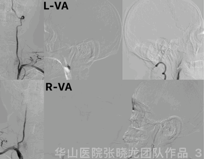
Figure 3 GIF. Bilateral vertebrate arteries angiogram showed bilateral posterior cerebral arteries P2 segment occluded and left posterior inferior cerebellar artery severe stenosis.
图 3 GIF. 双侧椎动脉造影证实双侧大脑后动脉P2以远闭塞,左侧小脑后下动脉重度狭窄。
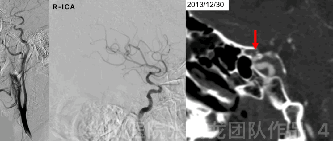
Figure 4 GIF. Right ophthalmic segment focal severe stenosis was noted. Dyna-CT reconstruction (9 years ago) revealed right ophthalmic segment severe stenosis with circumferential calcification (calcification ignored pre-operation).
图 4 GIF. 造影证实右侧颈内动脉眼段局部重度狭窄。9年前Dyna-CT重建提示右侧颈内动脉眼段重度狭窄伴环形钙化(术前未充分评估)。

Figure 5. The right ophthalmic segment stenosis did not aggravate compared with an angiogram nine years ago.
图 5 . 与9年前造影对比,右侧颈内动脉眼段狭窄未见加重。
The patient suffered from hypomnesia and slow reaction under antiplatelet therapy.
The right ICA ophthalmic segment stenosis and left ICA in-stent restenosis will be treated to increase perfusion to improve clinical symptoms.
However, circumferential calcification of the right ICA ophthalmic segment stenosis on the was ignored before the operation!
Left ICA in-stent restenosis, bilateral occluded posterior cerebral arteries, and left posterior inferior cerebellar artery severe stenosis formed due to unsatisfactory blood sugar control.
Systolic blood pressure should be strictly controlled if the stenosis was dilated satisfactorily.
抗血小板治疗下患者仍有记忆减退、反应迟钝。
通过治疗改善右侧颈内动脉眼段狭窄及左侧颈内动脉支架内再狭窄,增加脑灌注,缓解临床症状。
然而右侧颈内动脉眼段狭窄伴环形钙化,术前被忽视!
左侧颈内动脉支架内再狭窄、双侧大脑后动脉闭塞及左侧小脑后下动脉重度狭窄与血糖控制不佳有关。
狭窄段打开后术后需要严格控制收缩压。
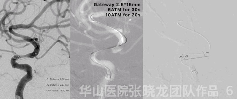
Figure 6 GIF. General heparinization was performed and 6F Envoy DA guiding catheter was placed near the right ICA cavernous sinus segment. Nimodipine 0.5ml was administered via guiding catheter to decrease blood pressure from 161/84mmHg to 135/64 mmHg. Gateway 2.5mm*15mm was positioned at the stenosis with the help pf a Synchro-II microwire. Dilated the balloon twice to 6 ATM for 30s and 10 ATM for 20s. The stenosis remained. Therefore retrieved the balloon and microwire.
图 6 GIF. 行全身肝素化。6F Envoy DA导引导管置于右侧颈内动脉近海绵窦段。经导引导管给予尼莫地平0.5ml将患者血压从161/84mmHg降至135/64 mmHg后,选用Gateway 2.5mm*15mm球囊在Synchro-II微导丝导引下至于右侧颈内动脉眼段狭窄段。以6ATM和10ATM分别充盈球囊30s和20s。复查造影狭窄段扩张不满意。遂撤出球囊和微导丝。
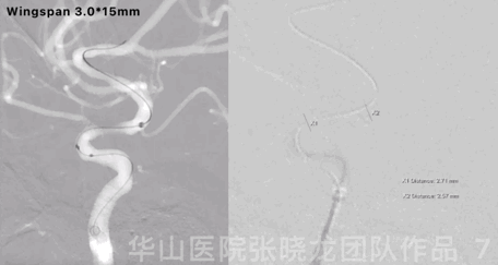
Figure 7 GIF. Deploy a Wingspan 3.0*15mm stent while the stenosis still dilated unsatisfactorily.
图 7 GIF. 于狭窄段释放Wingspan 3.0*15mm支架,复查造影狭窄段扩张仍不满意。
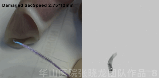
Figure 8 GIF. Attempted to advance SacSpeed 2.75*12mm balloon through the stent but failed. Retrieved the balloon and the SacSpeed 2.75*12mm damaged. Angiogram depicted the blood flow decreased and vasospasm occurred. Tirofiban 5ml and Nimodipine 0.5ml were administered.
图 8 GIF. 试图将SacSpeed 2.75*12mm球囊通过支架,反复尝试不能成功,遂撤出球囊。复查造影血流减慢,狭窄段血栓形成,血管痉挛。经导引导管给予替罗非班5ml和尼莫地平0.5ml。
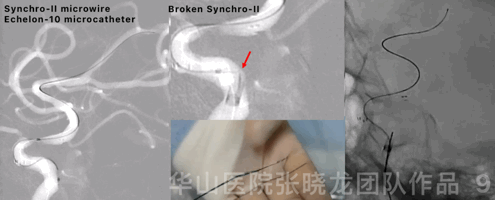
Figure 9 GIF. The distal part of the Synchro-II microwire broken due to repeated navigation of an Echelon-10 microcatheter. The broken microwire was retrieved along with the guiding catheter by a sharp-curved Transend-205 microwire (twisted and entangled) and a Gateway 2.5*9mm.
图 9 GIF. Echelon-10微导管反复尝试通过支架,反复尝试过程中Synchro-II微导丝远端断裂。Gateway 2.5*9mm球囊置于导引导管远端不出头,Transend-205微导丝远端塑急弯,微导丝远端成多个袢并缠绕前一断裂微导丝,连同导引导管一起撤出体外。
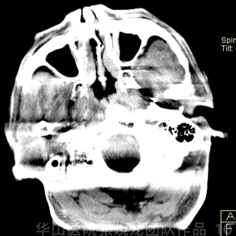
Figure 10 GIF. Dyna-CT showed no bleeding.
图 10 GIF. 复查Dyna-CT未见出血。
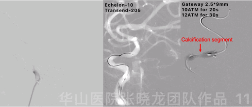
Figure 11 GIF. 6F Envoy DA was placed near the right ICA cavernous sinus segment and no bleeding was found from angiogram. Tirofiban 5ml was administered. An Echelon-10 microcatheter was navigated through the stent by a Transend-205 microwire. Dilated a Gateway 2.5*9mm balloon to 10ATM for 20s and 12ATM for 30s respectively in the stenosis segment.
图 11 GIF. 再次将6F Envoy DA导引导管置于近右侧颈内动脉海绵窦段,造影未见出血,予替罗非班5ml。Echelon-10 微导管在Transend-205微导丝导引通过支架。选用Gateway 2.5*9mm球囊分别在10ATM和12ATM下扩张20s和30s。
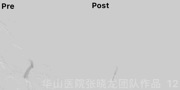
Figure 12 GIF. The stenosis did not improve after angioplasty.
图 12 GIF. 复查造影狭窄段扩张效果不佳。
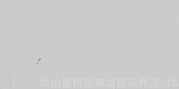
Figure 13. The right intracranial vessels were patent and no hemorrhage was observed.
图 13. 复查右侧颈内动脉造影颅内血管通畅,未见出血征象。
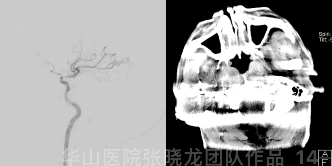
Figure 14 GIF. Rotational DSA showed the stenosis remained. Post-operative Dyna-CT did not show any bleeding.
图 14 GIF. 旋转DSA示狭窄仍然存在。术后Dyna-CT未见出血。

Figure 15. The rounded stiff calcification plaque aggravated from Dyna-CT reconstruction. The calcified plaque can be revealed clearly from the CT stead of fluoroscopy.The rounded stiff calcification plaque aggravated from Dyna-CT reconstruction. The calcified plaque can be revealed clearly from the CT stead of fluoroscopy.
图 15 GIF. 根据Dyna-CT重建图像,狭窄段钙化较9年前进展。钙化斑块在CT上显示清晰,透视下不能明确钙化。
Video 1. Another case with calcification plaque happened hemorrhage after balloon angioplasty, which should be paid attention to !
视频 1. 既往病例钙化斑块球囊扩张后出血,值得警惕!
1. The circumferentially stiff calcification plaque in the right ICA ophthalmic segment is a contraindication and should not be treated endovascularly.
Plaque calcification should be evaluated preoperatively from CT reconstruction instead of fluoroscopy.
Stenosis with calcified plaque can not be dilated well by the balloon while rupture and bleeding can occur after balloon angioplasty.
2. The Synchro-II standard microwire was easily broken due to repeated navigation of the microcatheter.
3. Broken microwire retrieve measure: Transend-205 with a sharp curve (several pigtail curves) twisted and entangled with the broken microwire.
4. The mild in-stent re-stenosis of the left ICA, was not the responsible lesion, which could be followed up.
1. 右侧颈内动脉眼段环形硬斑块,是血管成形术的禁忌。
术前需在CT重建上评估钙化斑块,透视下不能明确钙化。
狭窄伴钙化斑块,球囊扩张狭窄段困难,球囊扩张后会发生出血。
2. 微导管反复超选时Synchro-II微导丝容易折断。
3. 取出折断微导丝的方法:Transend-205远端塑急弯(多个猪尾巴样袢)缠绕断裂微导丝。
4. 左侧颈内动脉支架内轻度再狭窄,非本次发病的责任血管,可以继续随访。

点击或扫描上方二维码
查看更多“介入”内容
