2022年08月19日发布 | 912阅读

Immunity丨疾病相关的小胶质细胞和炎症相关的巨噬细胞的发育来源

爱大脑就AiBrain


撰稿 | AiBrain 内容团队

排版 | AiBrain 编辑团队


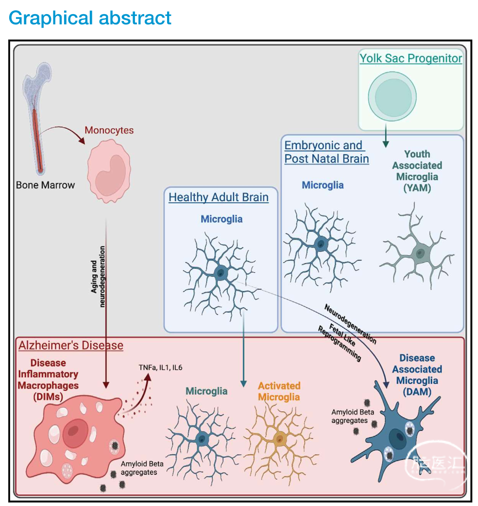
大脑中的巨噬细胞群包括实质小胶质细胞、边界相关的巨噬细胞(位于脑膜,脉络丛,血管周围的巨噬细胞)和单核细胞来源的细胞,它们共同维持着大脑的发育和生态平衡,然而它们也相互影响着衰老和神经退行性疾病的发病机制。然而关于大脑中的不同巨噬细胞群体在不同环境下的表型、定位和功能尚未得到解决。


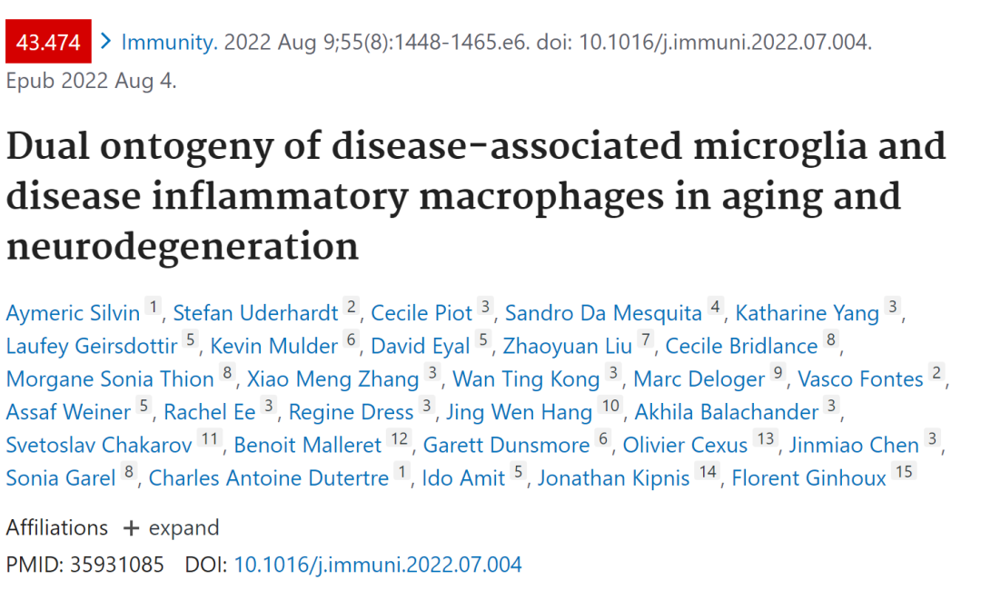
2022年8月9日,新加坡免疫协作组、上海交通大学基础医学院Florent Ginhoux研究团队在国际顶级期刊《Immunity》(IF=43.474)发表了文章。


研究者通过6个scRNA-seq大脑数据集的整合,生成了一个名为M-Verse的髓系图谱,以区分巨噬细胞群体的异质性。M-Verse显示了两个不同的巨噬细胞群体,DAM(疾病相关的小胶质细胞)和DIM(炎症相关的巨噬细胞),并发现DAM是胚胎来源,而DIM是TREM2非依赖的单核细胞来源,并揭示了DIM在疾病中的作用。

Florent Ginhoux

新加坡免疫学联网课题组长

上海市“海外高层次人才计划”

Ginhoux研究团队专注于了解骨髓细胞的起源、功能和稳态。其中包括树突状细胞(DCs)、单核细胞和巨噬细胞在组织稳态和免疫中发挥着关键而独特的作用;也致力于研究髓系细胞在疾病模型中的功能,探究治疗疾病新靶点。该研究团队旨在操纵这些细胞的潜在干预策略,并深入了解它们的起源和控制其内稳态的机制。已发表多篇颇具国际影响力的论文,其中包括Nature,Science,Immunity,Nature Immunology等40多篇高水平的国际期刊。

M-Verse,一个关于小鼠大脑

巨噬细胞异质性的全脑髓系图谱

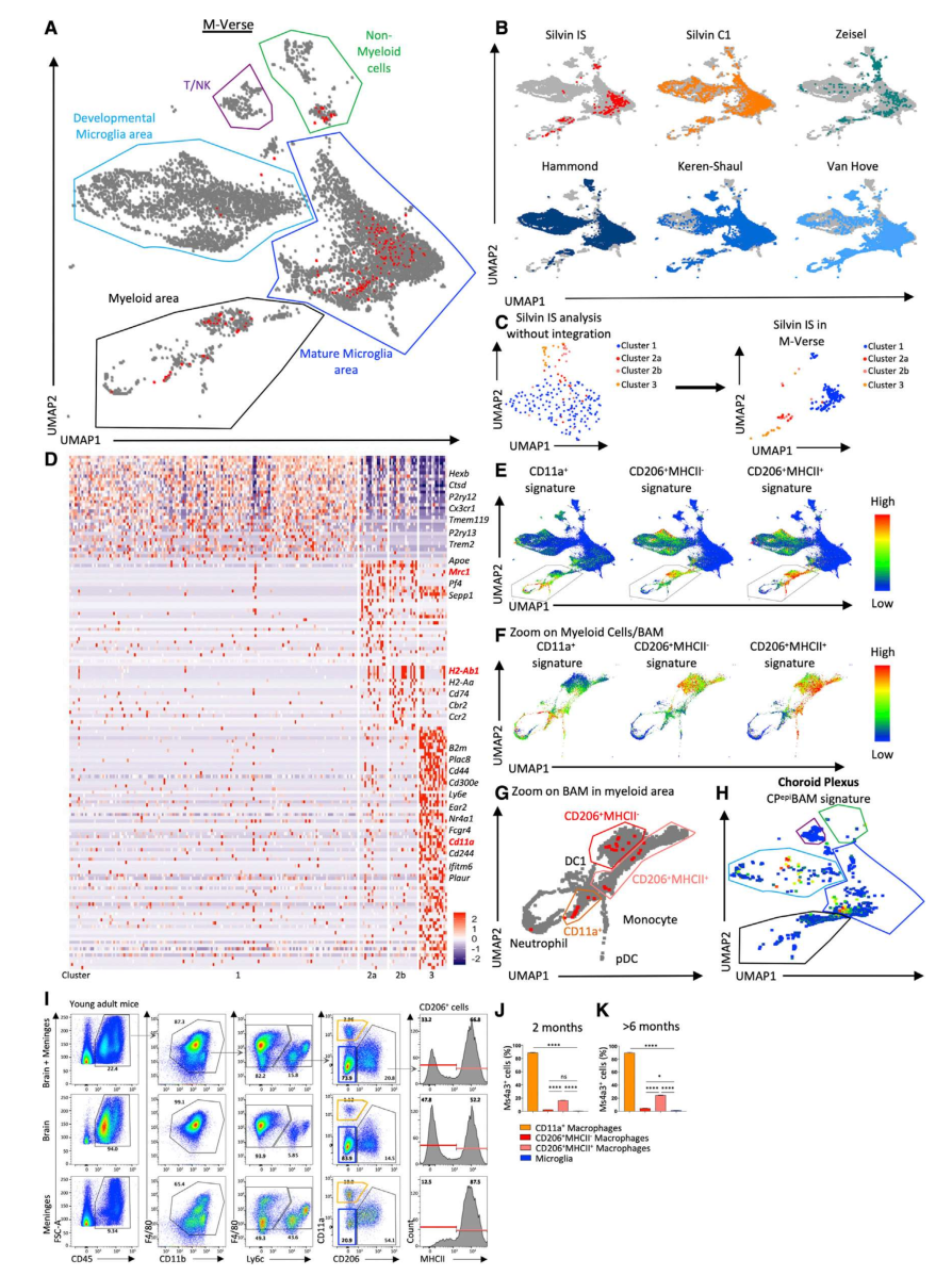
研究人员首先从成年小鼠的大脑中分离出骨髓细胞,并生成了一个索引排序(indexed-sorted, IS)scRNA-seq数据集,然后,对六个主要的已发表的scRNA-seq数据集(Hammond et al., 2019; Keren-Shaul et al., 2017; Van Hove et al., 2019; Zeisel et al., 2015)在发育期、成年期小鼠的大脑进行全局交叉比较,生成了小鼠大脑的全脑骨髓细胞图(称为"M-Verse"),研究人员从M-Verse(available online at https://macroverse.gustaveroussy.fr/2021_M-VERSE)中分析出小胶质细胞,CD206+BAMs(border-associated macrophage)的两个子集细胞群体,包括表达主要组织相容性II类分子(MHCII)的转录物和一个CD11a+单核细胞衍生的血管相关的亚细胞群体。


Figure 1. M-Verse, a global consensus of murine brain macrophage heterogeneity

(A) Six scRNA-seq dataset integration (186,026 cells): M-Verse. Red dots correspond to Silvin indexed-sort (IS) cells. (B) M-Verse split by individual datasets. In gray, cells from other datasets; in color, cells of respective dataset. (C) Silvin IS scRNA-seq dataset UMAP plot (left panel). M-Verse representation of Silvin IS dataset (right panel). (D) Cluster DEG heatmap, identifying putative signatures of microglia (1), CD206+ (Mrc1) MHCII+ (H2-Ab1) (2b), CD206+ MHCII (2a), and CD11a+ macrophages (3). (E) Projection of gene signatures of BAM clusters established in (D) on M-Verse. (F) Projection of gene signatures of BAM clusters established in (D) on the M-Verse myeloid cell/BAM area. (G) BAM regions among M-Verse based on (F) and localization of Silvin IS cells (in red). (H) CPepiBAM signature (Van Hove) projected on Van Hove cells within the M-Verse. (I) Gating strategy for microglia, CD206+MHCII+ macrophages, CD206+ MHCII macrophages, and CD11a+ macrophages identified by flow cytometry of C57BL/6 young adult mouse brains (with meninges), meninges, and brain alone (n = 3). (J) Macrophage ontogeny in 2-month-old fate mapping mouse (n > 3). (K) Macrophage ontogeny in >6-month-old fate mapping mouse (n > 3). One-way ANOVA; *p value < 0.05, ****p value < 0.0001.

△上下滑动查看△


M-Verse数据库揭示

BAM的起源和DAM的异质性

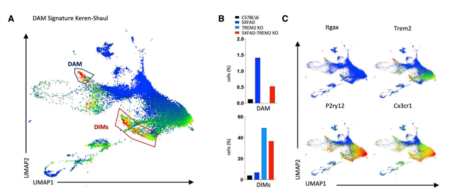
中枢神经系统除了小胶质细胞,还包含边界相关巨噬细胞(BAM, border associated macrophages ),它们位于中枢神经系统的脑膜、脉络膜丛和血管周围空间。基于M-Verse和标志性DEG编码蛋白的表达,研究者设计了一个严格的流式细胞仪门控策略,标记不同的巨噬细胞群。这些标记基因和蛋白包括Mrc1(编码CD206)、Itgal(编码CD11a)和H2-Ab1(编码MHCII)。将基因Ms4a3可用于谱系单核细胞谱系追踪,研究者发现不到1%的小胶质细胞被标记,而超过87%的CD11a+巨噬细胞被标记,这表明后者主要来自单核细胞。其中更多数量CD206+MHCII+巨噬细胞来源于单核细胞。这表明不同区域的BAM来源不同。DAM是大脑中一类重要的巨噬细胞亚群,被认为在AD疾病中发挥着保护功能,在AD患者大脑中也能够检测到DAM。另一项研究表明,DAM也会在AD小鼠的大脑中积累,但其作用是否具有保护作用仍存在争议。因此研究者利用M-Verse对DAM亚群特性进行深入分析,发现高度表达DAM基因的细胞位于两个不同的区域:一些位于胚胎时期来源的小胶质细胞区域(DAM);另一些位于在成熟的小胶质细胞区域内,因其高表达炎症相关的基因,将其定义为疾病炎症相关巨噬细胞(disease inflammatory macrophages, DIMs)


Figure 2. DAM signature includes two distinct cell populations.

(A) Heatmap of DAM signature determined by Keren-Shaul et al., projected onto Keren-Shaul dataset with M-Verse coordinates, revealing two regions enriched with the Keren-Shaul DAM signature, named DAM and DIMs. (B) DAM and DIM percentage among total cells from Keren-Shaul mouse AD models. (C) Relative RNA expression heatmap of specific DAM markers described by Keren-Shaul et al., projected on Keren-Shaul dataset with M-Verse coordinates.

△上下滑动查看△


Figure 3. DIMs increase in frequency during aging and in AD murine models. 

(D) Venn diagram showing common and unique DEGs expressed at a higher level in DIMs compared with DAM in Hammond, Keren-Shaul, and Van Hove datasets and establishment of a DIM-conserved gene signature. (E) DIM-conserved signature heatmap among all the M-Verse. (F) Pathways associated with DAM or DIM signatures.

△上下滑动查看△


DAM与YAM的功能性差异

接下来,研究者继续探究DAM在其他数据库中(Keren-Shaul and Van Hove overlaid)是否与其他细胞共同表达一些基因,其中发现八个基因特异性表达在DAM细胞上的基因(Dkk2, Fabp5, Gpnmb, Igf1, Itgax, Mamdc2, Spp1, Gm1673),数据分析还发现成年DAM与小鼠的发育期(胎儿期, E和产后早期,P)的小胶质细胞有着共同的基因表达特征(Dkk2, Gpnmb, Igf1, Itgax, Spp1),包括整合素Itgax(CD11c)的表达。由于神经退行性疾病中发现的DAM保守基因与野生型小鼠胚胎和新生儿大脑中的一组细胞非常相似,基于这些观察结果,将CD11c+ P7小胶质细胞称为青年相关小胶质细胞(YAM, youth associated microglia)。YAM 和DAM 均表达大量与吞噬、自噬和线粒体代谢相关的基因。尽管YAM和DAM有共同的基因表达模式,但前者在胚胎发育和出生后早期自然出现,代表了与大脑发育有关的小胶质细胞群,而DAM则相反,在神经变性的情况下,DAM会大量积累,可能参与了恢复大脑的平衡状态的过程。


Figure 4. DIMs are monocyte derived, whereas DAM are embryonic derived

(A) Schematic overview of irradiated mice and cell populations sorted from 5-month-old mice brain. (B) Gating strategy to sort corresponding populations (top panel). Visualization of the three cell populations along the gating strategy (bottom panel). (C) Histogram percentages of sub-populations among all brain macrophages (n = 3). (D) GSEA enrichment analysis of DIM signature established in the M-Verse among the three brain macrophage populations sorted from irradiated mice. (E) Flow cytometry measurement of F4/80, CD11b, CD83, P2RY12, MHCII, and CD11c expression by indicated populations. (F) Gating strategy to delineate brain DIMs among brain cells from Ms4a3creRosaTomato 2- and >6-month-old mice. Representative dot plot. (G) Histogram showing DIM percentage among all brain macrophages (n > 5). (H) Zoom on the three areas enriched in Ms4a3-TdT+ cells in >6-month-old mouse brain, representative of 3 mice. (I) Representative gating strategy for the identification of DIMs (red) and DAM (blue) in C57BL/6, 5XFAD, TREM2-/-, and 5XFAD-TREM2-/-12-month-old mice. One-way ANOVA *p value < 0.05. (J) Histogram showing DIM and DAM percentage among all macrophages in C57BL/6, 5XFAD, TREM2-/-, and 5XFAD-TREM2-/-mice (12 months old; one-way ANOVA *p value < 0.05). (K) Histogram showing Ms4a3-TdT expression among DIMs and DAM from 12-month-old C57BL/6 and 5XFAD mice (representative n = 4). (L) Histogram showing Ms4a3-TdT+ percentage among DIMs and DAM in 12-month-old 5XFAD mice (n = 3). (M) Histogram showing CD45, CD11b, F4/80, Ly6C, CD83, CX3CR1, CD11c, MHCII, CD206, and CD11a expression among DAM, DIMs, and microglia (12-month-old 5XFAD mice). Mann-Whitney test **p value < 0.01, ****p value < 0.0001.

△上下滑动查看△


DIMs起源于外周浸润的巨噬细胞

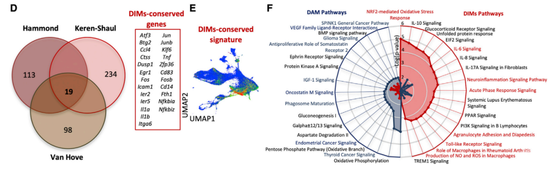
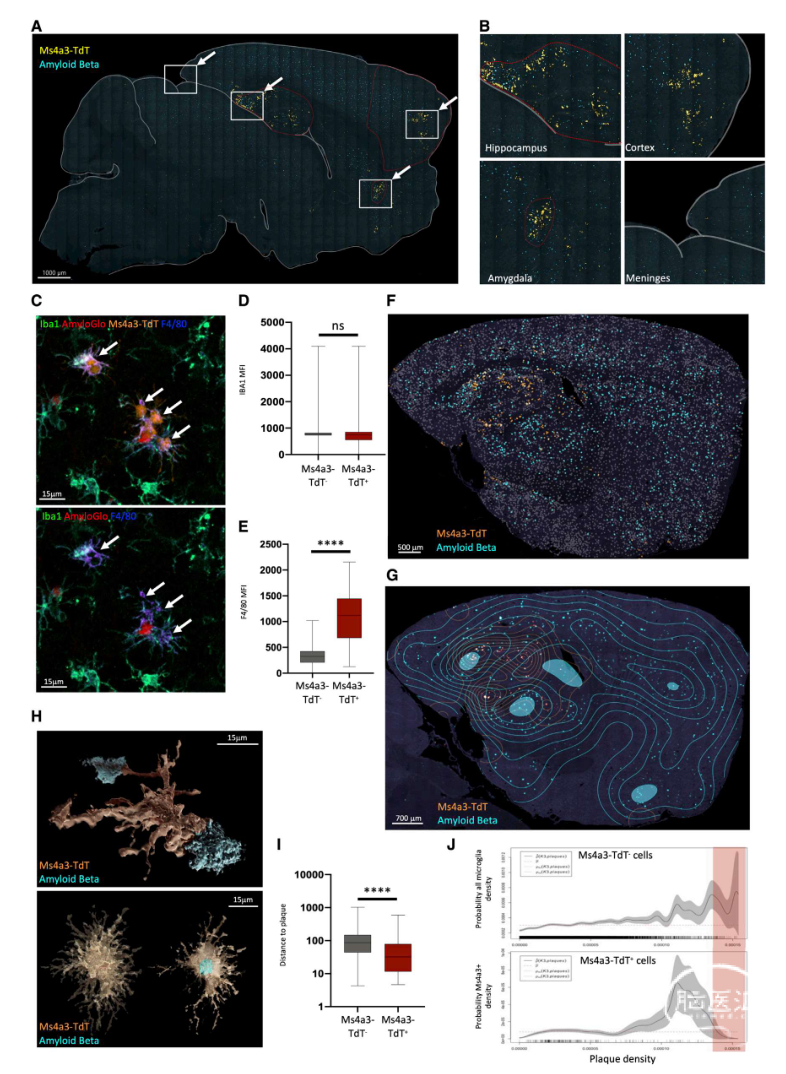
然后,研究者也探究另一种细胞群体的特征-DIMs(disease inflammatory macrophages, DIMs),DIMs出现在小鼠出生之后,随着衰老的进行,逐渐增加。在AD疾病环境中,大脑处于炎症状态,炎症因子水平的增加,增加DIMs细胞的积累。研究者利用经过辐照的小鼠模型,观察到外周细胞对大脑的浸润,采用细胞命运谱系追踪发现DIMs来源外周巨噬细胞,且不依赖于TREM2。而在大脑炎症或神经退行性疾病状态下,DIM来源于高表达CD83, F4/80, Ms4a3的单核细胞,且主要分布在海马,丘脑和皮层区域,因此DIM是单核细胞起源,且不依赖于TREM2,并随着衰老和炎症状态大量增加。


DIM在AD疾病中的作用

为进一步了解DIM在疾病中的作用,研究者采用4种转基因小鼠(C57BL/6-Ms4a3CreRosaTomato、5XFAD-Ms4a3CreRosaTomato、C57BL/6-TREM2-/- and 5XFAD-TREM2-/-模型小鼠,揭示了DAM和DIMs是两个在本体发育上不同的细胞系。可以通过F4/80、CD83、CX3CR1、CD11c和CD206的表达量来区分,并表现出对TREM2不同的依赖性(DAM依赖于TREM2信号途径,而DIM不依赖于TREM2途径)。研究人员通过免疫荧光发现DIM细胞群主要聚集在阿尔兹海默症模型小鼠的海马、脑膜、基底外侧杏仁核和皮质,并位于β 淀粉样蛋白聚集物附近,且在AD患者脑中也发现CD83+ TNF-a+ DIMs出现在软脑膜中。由此可见,在小鼠中发现的DIMs(或许DAM和YAM)scRNA-seq图谱可以扩展到人类疾病中的研究。


Figure 5. DIM clustering in 12-month-old 5XFAD mice specific brain regions

(A) Full brain slice imaging of Ms4a3-TdT+ and Ab aggregates. White square, area enriched in Ms4a3-TdT+ cells. (B) Areas enriched in Ms4a3-TdT+ cells from (A),(C) Iba1 and F4/80 expression by Ms4a3-TdT+ cells and amyloGlo. (D) Iba1 expression intensity among microglia and Ms4a3-TdT+ cells (n = 3). (E) Intensity of F4/80 expression among microglia and Ms4a3-TdT+ cells (n = 3). (F) Analysis of the distribution of Ab aggregates and Ms4a3-TdT+ cells. (G) Density analysis of the distribution of Ab aggregates and Ms4a3-TdT+ cells. (H) 3D reconstruction of Ms4a3-TdT+ cells interacting with Ab aggregates without (top panel) or with (bottom panel) transparency. (I) Histogram of the distance-to-plaque (mm) from microglia or Ms4a3-TdT+ cells (n = 3). (J) Microglia or Ms4a3-TdT+ cell density analysis depending on plaque density. Mann-Whitney test ****p value < 0.0001.

△上下滑动查看△


总结:

本研究通过整合六个不同的数据集生成M-Verse,创建了一个横跨从胚胎发育到衰老和神经退化的免疫细胞图谱,揭示了小胶质细胞和巨噬细胞亚群的复杂性,进一步探究DAM异质性,揭示出两个本体发育和功能上不同的细胞系(DAM和DIMs)。胚胎来源的DAM只出现在神经变性的背景下,而在正常衰老过程中并不明显,而单核细胞衍生的DIMs随着年龄的增长,及在炎症和AD神经变性过程中逐渐积累。


点评:

本研究揭示了两个本体发育和功能上不同的细胞系(DAM和DIMs),且在AD疾病中的作用,然而有关小胶质细胞异质性的研究中,还发现了YAM(youth associated macrophage)PAM(proliferative-region associated microglia)、ATM(axon track microglia) 进一步的研究将直接评估YAM在胚胎发育中的作用和DAM在神经退行性疾病中的作用,以及其他细胞亚群之间的不同功能的比较。总之M-Verse数据库使我们能够交叉比较几项研究并解决有关各种巨噬细胞群的性质和本体发育的重要问题,及在不同的数据集中的各种巨噬细胞群的性质和本体发育的重要问题。


摘要:

Brain macrophage populations include parenchymal microglia, border-associated macrophages, and recruited monocyte-derived cells; together, they control brain development and homeostasis but are also implicated in aging pathogenesis and neurodegeneration. The phenotypes, localization, and functions of each population in different contexts have yet to be resolved. We generated a murine brain myeloid scRNA-seq integration to systematically delineate brain macrophage populations. We show that the previously identified disease-associated microglia (DAM) population detected in murine Alzheimer’s disease models actually comprises two ontogenetically and functionally distinct cell lineages: embryonically derived triggering receptor expressed on myeloid cells 2 (TREM2)-dependent DAM expressing a neuroprotective signature and monocyte-derived TREM2-expressing disease inflammatory macrophages (DIMs) accumulating in the brain during aging. These two distinct populations appear to also be conserved in the human brain. Herein, we generate an ontogeny-resolved model of brain myeloid cell heterogeneity in development, homeostasis, and disease and identify cellular targets for the treatment of neurodegeneration.


PMID: 35931085

DOI: 10.1016/j.immuni.2022.07.004

全文链接:

https://pubmed.ncbi.nlm.nih.gov/35931085/


往期精彩回顾


声明:脑医汇旗下神外资讯、神介资讯、脑医咨询、AiBrain所发表内容之知识产权为脑医汇及主办方、原作者等相关权利人所有。未经许可,禁止进行转载、摘编、复制、裁切、录制等。经许可授权使用,亦须注明来源。欢迎转发、分享。


最新评论
发表你的评论
发表你的评论
来自于专栏
相关临床评分小工具
关键词搜索