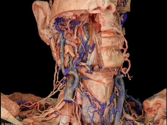
▼这是上颌动脉
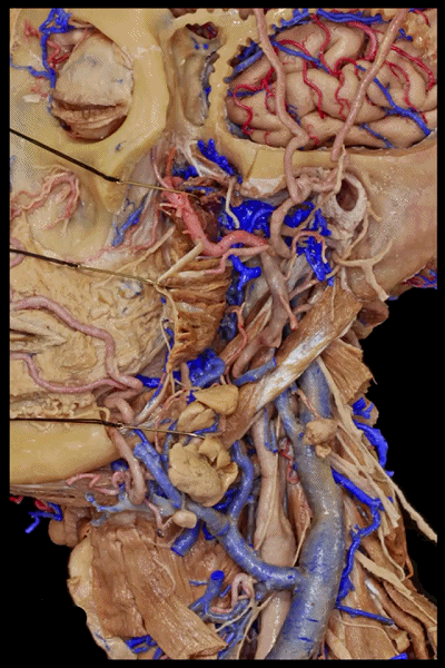
▼这是颞浅动脉
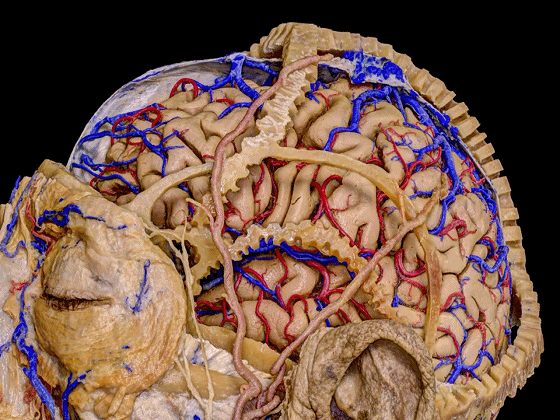
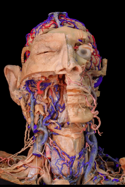
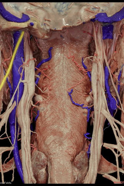
▼来看这一标本的侧面观,去除了下颌骨、茎突肌、二腹肌。显露出口腔
So this is a lateral view of a specimen with the mandible, the styloid and digastric muscles removed. The oral cavity has been opened,

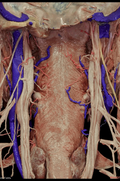
▼我们来看一下颈外动脉的主要分支:这是甲状腺上动脉(下图),可用于血管搭桥。
and the main branches of the external carotid artery can bedistinguished, the superior thyroid artery, which can be used as a vascular graft,

▼这是面动脉,已被切断
the facial which has been cut here,

▼这是舌动脉的终末支
the lingual, we see the terminal branch of the lingualartery,

▼这是枕动脉
▼这是上颌动脉,行于颞下窝。
and the internal maxillary artery which runs in the infratemporal fossa.

▼这里显示颈外动脉的两终末支:上颌动脉、颞浅动脉,常规用于搭桥手术。
We see here the two terminal branches of the external carotid artery, maxillary artery, and superficial temporal artery which isused...commonly used for bypass surgery.
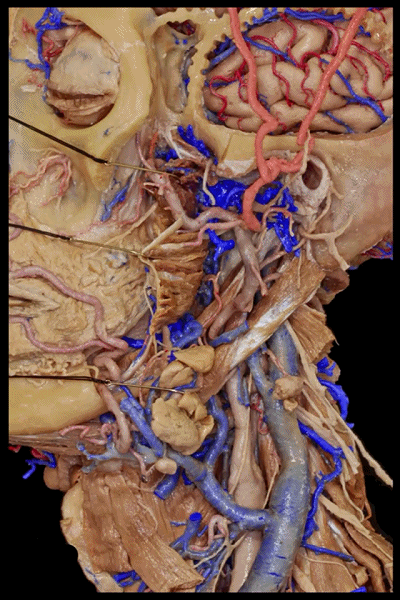
▼这是其与面神经颞支(下图)的关系。
And here is relationships with the frontalis branch of thefacial nerve.
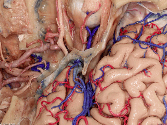
▼这是上方的 经颅手术入路 视角,可见眼眶,外侧裂。
This surgical position, position from above, we can see here the orbit, the Sylvian fissure.
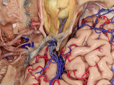
▼我们磨除部分中颅底骨质,显露出上颌动脉(下图),可将其与大脑中动脉行吻合术,故可用于搭桥。
We have drilled part of the middle fossa floor, and we see the internal maxillary artery, that can be anasomosed to the middle cerebral artery in supposing a vascular graft.
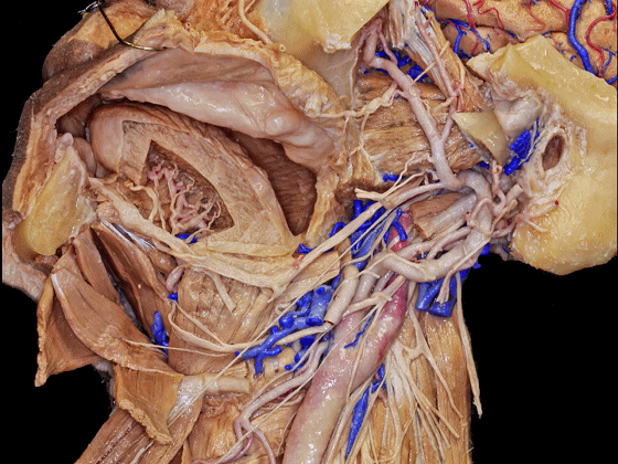
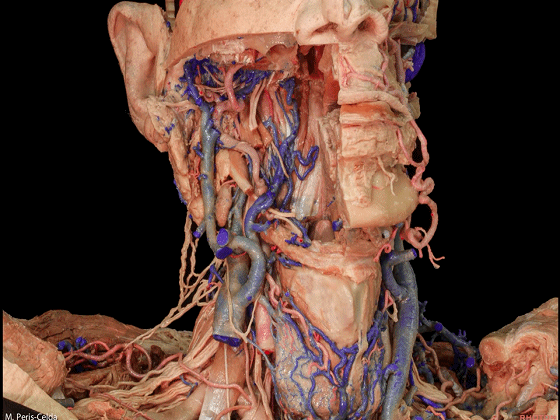
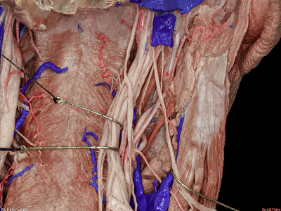
▼颈前三角(下图)也常在颈椎手术的入路中打开,因此需掌握其神经血管的解剖毗邻。
So the anterior triangle of the neck is also entered in approaches to the cervical spine, and its vascular and neural relationships are very importantto minimize complications in this area.
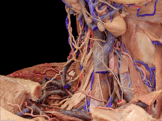
▼这是上方视角,右侧锁骨已被切除,可见如下重要解剖结构。中线部位可见甲状腺(下图)
So in this view from above, after removal of the right clavicle, we can have the important references of this anatomy. So in midline we see the thyroid gland,
▼大血管包括颈动脉、颈内静脉已被部分切除(下图)
then we see the great vessels, the carotid artery, and theinternal jugular vein that have been divided,
▼这是迷走神经,走行于上述两大血管后方。
the 10th nerve, that goes behind these two vessels.

▼这是前斜角肌
We see the anterior scalenus muscle
▼这是膈神经
▼在前斜角肌后方,可见臂丛
Behind this, we see the brachial plexus,
▼上方是颈丛
the cervical plexus above

▼这是副神经
the accessory nerve here.
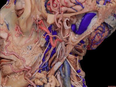
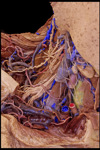
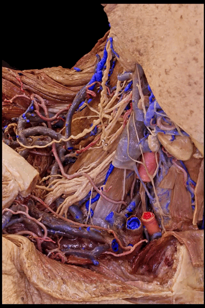
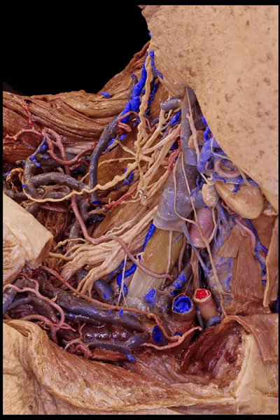
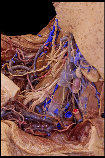
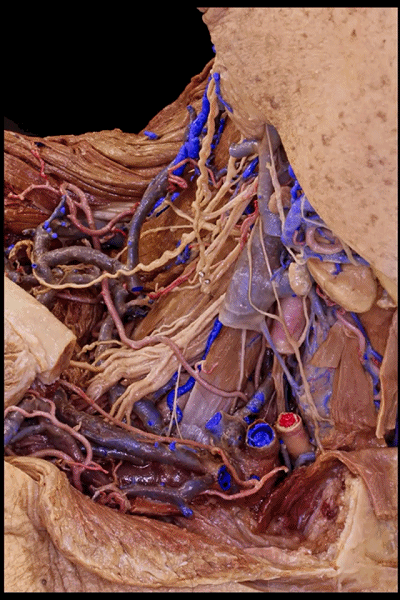
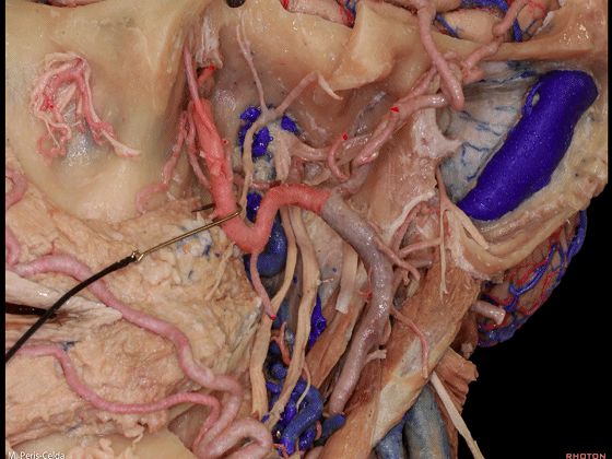
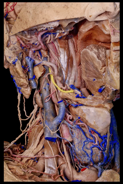
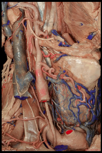
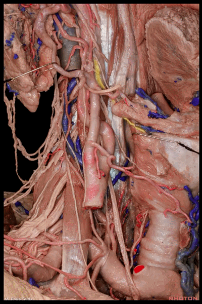
▼这里,我们切除右侧下颌骨和上颌骨,以暴露部分颞下窝。部分翼状肌已被切除。可见咽旁间隙,以及椎前间隙。这是上颌动脉(下图)。
So, removal of the right mandible and maxilla shows part of the infratemporal fossa.Part of the pterygoid muscles have been removed.We can see the parapharyngeal space,and the spanvertebral space. This is the internal maxillary artery.
▼这是突出的寰椎前结节(下图),位于椎前间隙内,口腔后方。
The anterior tubercle of the atlas protrudes right here, in the spanvertebral space, and behind the oral cavity,

▼寰椎前结节为颈长肌(下图)的上附着处。颈长肌位于中线。
and serves as a superior attachment of the longus collimuscle.which is at midline.
▼头长肌(下图)恰位于颈长肌外侧。
Longus capitus muscle is going to be right lateral to the longus colli muscle,
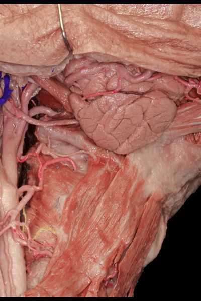
▼咽旁间隙由茎突筋膜(下图)分成两部,茎突筋膜与茎突肌群相延续,此处已被切除。
The parapharyngeal space is divided by the styloid fascia, which is continued with the styloid muscles divided here,

▼咽旁间隙分为茎突前部和茎突后部。
in span- and poststyloid compartments
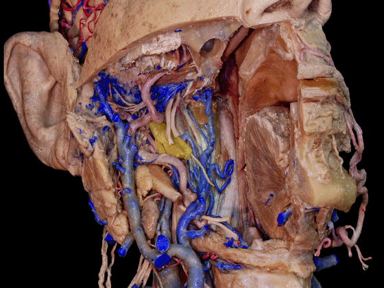
▼茎突前部(下图)位于翼内肌内侧,其内充满脂肪,有咽升动脉分支、面动脉、以及舌咽神经分支通过。
The spanstyloid compartment medial to the medial pterygoid muscle, is filled with fat that contains branches of the ascendingpharyngeal or facial arteries, and branches from the glossopharyngeal nerve.
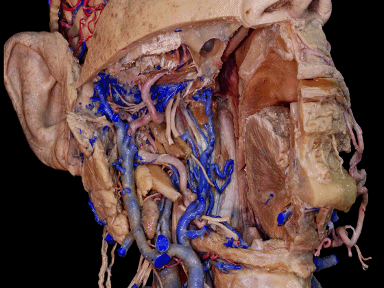
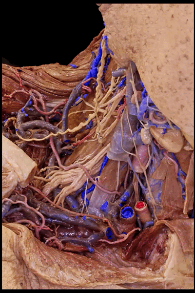
▼这显示的是茎突筋膜(左侧),翼状肌已被切除,其位于三叉神经下颌支的后方,以及上颌动脉的后方。
This is a view of the styloid fascia,after removal of the pterygoid muscles,behind the branches of the 3rd division of the trigeminalnerve,and internal maxillary artery.

▼这是三叉神经下颌支
▼这是上颌动脉
▼茎突后部(下图)又称为岩下间隙,位于岩骨的下方,茎突筋膜(下图)的后方,乳突尖的内侧。
The poststyloid compartment is also called infrapetrosal, is posterior to the styloid fascia, inferior to the petrous bone, and medial to mastoid tip.
▼走行于茎突后部内的重要结构包括:颈内动脉颈段的上部(下图)、颈内静脉(下图)、舌咽神经、迷走神经、副神经、舌下神经。
Some of the important structures in the area are the superior aspect of the cervical segment of theinternal carotid artery,jugular vein,9th,10th,11th,and 12th cranial nerves.

▼这是舌咽神经,其位于颈内动脉前方
anterior to the internal carotid,

▼这是迷走神经,其位于颈内动脉后方
behind the internal carotid,

▼这是副神经
▼副神经跨过颈内静脉,位于静脉前方,但有时也可位于其后方。
and 11th is crossing the internal jugular vein, anterior, but sometimes is posterior to it.
▼这是舌下神经

▼舌下神经绕行于颈内和颈外动脉,位于舌骨上方,二腹肌下方。
The 12th cranial nerve surrounds both carotid arteries, above the hyoid bone, and below the digastric muscle.
▼胸锁乳突肌动脉(下图)是枕动脉(下图)的分支,位于舌下神经(下图)上方。切断胸锁乳突肌动脉有助于将舌下神经移向上方,以暴露颈动脉。
Dividing the sternocleidomastoid artery,which is branch of the occipital artery, just above the hypoglossal nerve, aids in safely retracting the nerve superiorly in approaches to the carotid artery.

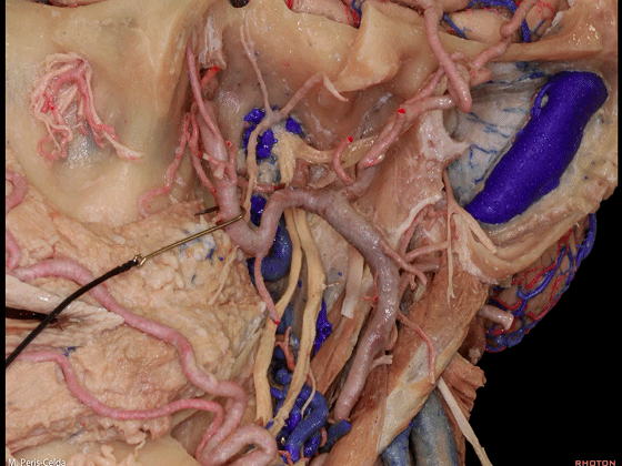
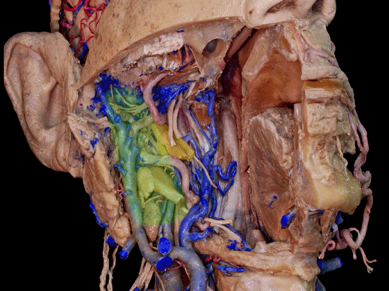
▼从后面来暴露茎突后部,这里已去除颈椎,可见交感神经节(下图)及交感干,位于该区域的最内侧部。
So the poststyloid compartment from behind after removal of the cervical spine, reveals that the sympathetic ganglion and the trunk are the most medial structures in this area.
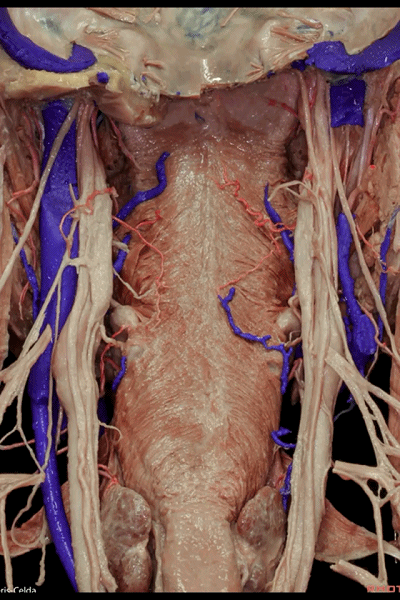
▼这是交感干
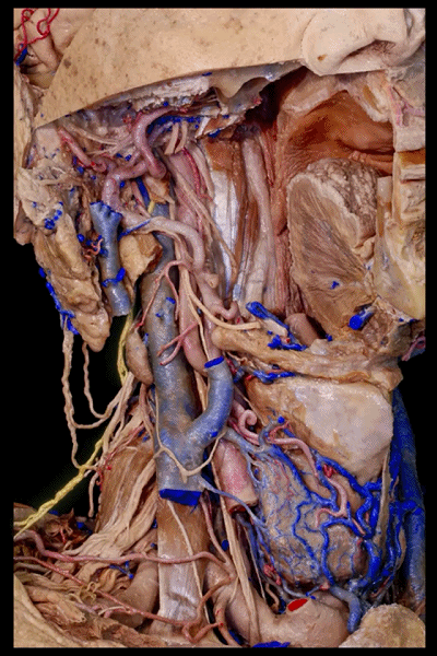
▼迷走神经走行于颈内静脉和颈动脉之间。
Then we have here the 10th nerve running in between the jugular vein and carotid artery.

▼在其后方为颈神经(下图)。
Behind them, these are the cervical spinal nerves.
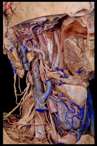
▼副神经在这一侧位于颈内静脉的后方
The accessory nerve here running behind the jugular vein on the side,
▼而在这一侧,副神经位于颈内静脉的前方
and anterior to it on this side.
▼换成侧后方的角度,可见舌下神经从后方包绕颈内动脉和颈外动脉,随后前行支配舌。
And in a lateral view of the same specimen, the hypoglossal nerve which is posterior surrounds the internal and external carotid arteries, and goes towards the tongue.

▼这是颈袢(ansa cervicalis)
And this is the ansa cervicalis,that we saw spanviously.

▼这是副神经
This...they are accessory nerve

▼这是舌咽神经
the glossopharyngeal nerve here
▼这是颈上交感神经节和交感干,位于最内侧。
and the superior cervical ganglion and sympathetic trunk, which are the most medial structures.

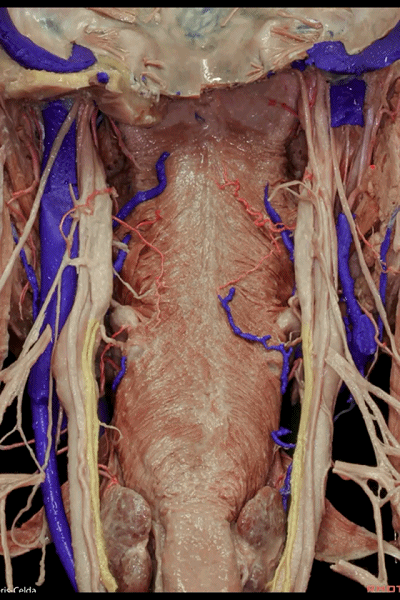
▼有两条重要的神经,会出现在暴露椎前间隙和颈椎的手术入路中,即喉上神经、喉下神经,均为迷走神经的分支。它们从外侧行至内侧。
So there are two important nerves in the approach of thespanvertebral space and cervical spine, that cross from lateral to medial, and are the superior and inferior laryngeal nerves, which are branches of the 10th cranial nerve.
▼喉上神经又分为两支:内支、外支。
这是内支(下图),内支在上,位于舌骨(下图)下方,穿经甲状舌骨膜。
So the superior laryngeal nerve is divided in two: this is the internal branch, The internal branch is superior and can be found inferior to the hyoid, piercing the thyrohyoid membrane.

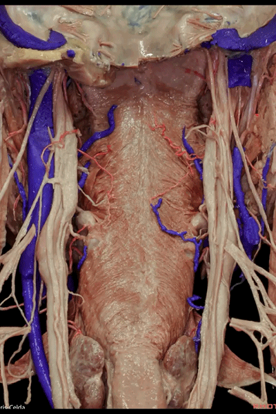
▼喉上神经 内支为感觉支。位于舌骨(下图)下方。因此,单侧病变若累及喉上神经的内支,将导致喉部的异物感,而双侧病变则可导致误咽。
and the internal branch has a sensory function. So the function of the internal branch we see here... the internal branch below the hyoid,So lesions of the...unilateral lesions of the internal branch of the superior laryngeal nerve produce a feeling of foreign body in the throat, and bilaterally, they may result in aspiration.
▼喉上神经 外支(下图)在下方,较细,位于甲状腺上动脉(下图)的深面。
And theexternal branch is thinner, is inferior, and can be found deeper to the superior thyroid artery.
▼喉上神经 外支支配环甲肌(下图)。累及单侧外支的病变,可无明显症状,或可导致音色的改变或发音的易疲劳性。
It innervates the cricothyroid muscle,Unilateral lesions of the external branch may be not advertent or the patient may have an impaired ability to produce acutesounds and his voice fatigability.

▼另一重要神经即喉下神经(又称喉返神经 下图),在右侧,其绕行于锁骨下动脉(下图),在左侧,其绕行于主动脉弓。
And another nerve is the inferior laryngeal nerve orrecurrens, which loops around the subclavian artery on the right side, and around the aortic arch on the left side.

▼喉下神经其进入喉的部位 位于喉的下部,食管和气管之间
It enters the larynx through this point inferior and between the esophagus and the trachea

▼喉下神经负责喉上神经外支所支配肌肉以外的所有喉肌。即除环甲肌(下图)以外的所有喉肌。累及单侧喉下神经的病变,也可以无症状或出现声音嘶哑或呼吸声。双侧病变则可很严重而出现喘鸣。
and innervates all the muscles that are not innervated bythe external laryngeal nerve. So,all the muscles except the cricothyroid muscle. and the unilateral damage of the inferior laryngeal nerve, may be asymptomatic or the patient may have most commonly a hoarse or breathy voice. And bilateral lesions may be very serious and may causestridor.

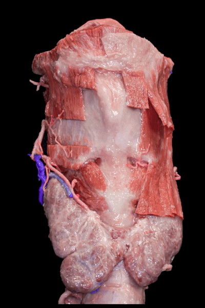
▼这是喉的前面观,可见舌骨(下图)、甲状软骨、环状软骨、甲状腺。
So this is a front view of larynx, with the hyoid bone, the thyroid cartilage, the cricoid cartilage, and the thyroid gland.

▼这是甲状软骨
the thyroid cartilage

▼这是环状软骨

▼这是甲状腺
声明:脑医汇旗下神外资讯、神介资讯、脑医咨询、AiBrain所发表内容之知识产权为脑医汇及主办方、原作者等相关权利人所有。未经许可,禁止进行转载、摘编、复制、裁切、录制等。经许可授权使用,亦须注明来源。欢迎转发、分享。


