Case Review
History
Video 1. Angiography shows two ophthalmic artery aneurysms with ipsilateral ICA dissection. One of the ophthalmic aneurysm is on the course of oculomotor nerve. 血管造影显示两枚颈眼动脉瘤伴同侧颈内动脉夹层。其中一枚颈眼动脉瘤位于动眼神经旁。
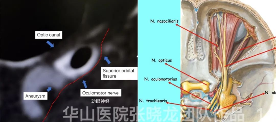
Figure 1. Ptosis was due to the left oculomotor nerve comspanssed by the aneurysm. 上睑下垂考虑为左侧动眼神经受动脉瘤压迫所致。
1
Strategy
Two aneurysms located at the ophthalmic segment of the left ICA.
Stent-assisted coiling – it may be difficult to embolize the second aneurysm after stent deployment. FD stent can simplify the procedure.
2.The ICA dissection can be treated with carotid stent implantation. During the procedure, the guiding catheter should be first navigated into the cavernous segment of the ICA for FD deployment. Then retrieve the guiding catheter and deploy the carotid stent.
支架辅助栓塞——支架释放后很难栓塞第二枚动脉瘤。 血流导向支架可简化操作。
2.颈内动脉夹层行颈动脉支架植入术。术中应先将导引导管送至颈内动脉海绵窦段来以便更好释放血流。然后撤出导引导管并放置颈动脉支架。
2
Operation
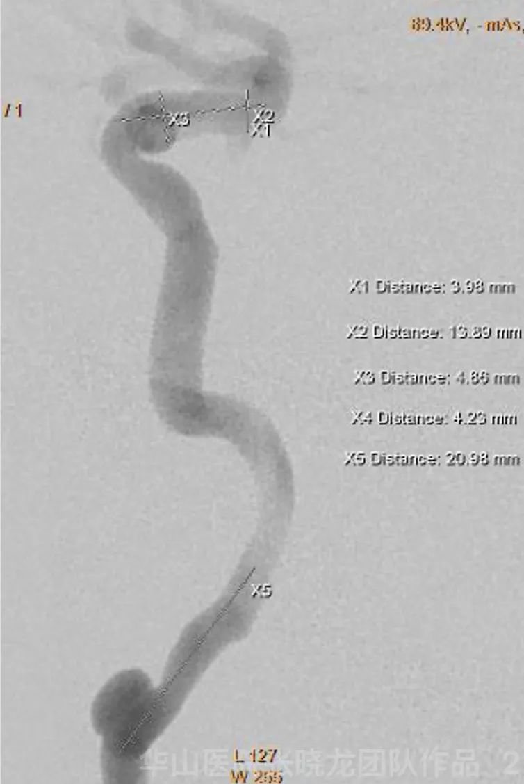
Figure 2. General Heparinization was administrated at the beginning of the operation. 手术开始时行全身肝素化。
Figure 3 GIF. 6F 90cm sheath. 6F 115cm Navien was placed at the cavernous segment of the left ICA. 采用6F 90cm的鞘。6F 115cm Navien导管置于左侧颈内动脉海绵窦段。
Figure 4 GIF. Marksman microcatheter was navigated to the left M1 guided by Synchro II standard microwire. Marksman微导管在Synchro Il微导丝引导下送至左侧大脑中动脉M1段。
Video 2. Pipeline 4.0*20mm was deployed covering two ophthalmic artery aneurysms. 释放Pipeline 4.0*20mm支架覆盖两枚颈眼动脉瘤。
Figure 5 GIF. Precise 6*40mm stent was deployed for remodeling the dissecting segment of the left ICA. 用Precise 6*40mm支架来重塑左侧颈内动脉夹层段。
Figure 6 GIF. Post-operation angiography shows the contrast medium retention in the aneurysm sac and the dissection. 术后血管造影显示动脉瘤腔及夹层内造影剂滞留。
Figure 7 GIF. 3D reconstruction shows the well-deployment of the Pipeline stent. 三维重建显示Pipeline支架放置良好。
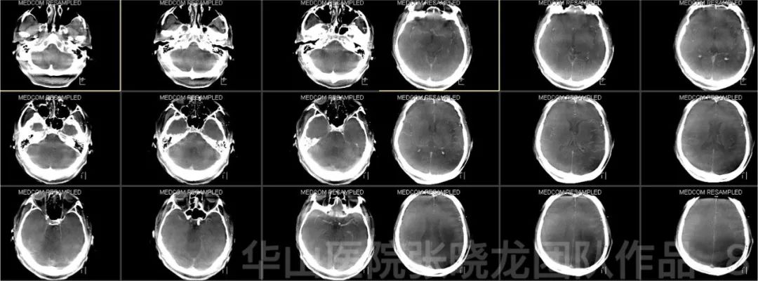
Figure 8. Post-operative Dyna CT shows no intracranial hemorrhage or infraction. 术后Dyna CT未见颅内出血或脑梗死。
3
Post-operation
• No new neurologic deficit.
4
Follow up
3-month follow up 三个月随访
• Dual antiplatelet therapy was used for one month.
• Therapy was stopped by the patient himself.
• 左侧上睑下垂术后2个月恢复。

Figure 9 GIF. 3-month follow up angiography shows the complete occlusion of two ophthalmic artery aneurysms with patent parent artery and mild stenosis proximal to the dissection. 三个月随访血管造影显示两枚颈眼动脉瘤完全栓塞,载瘤动脉通畅,夹层近端有轻度狭窄。
5
Summary
• In the case with two ophthalmic artery aneurysms, flow divert treatment is recommended for simplifying the procedure, which is also beneficial to oculomotor nerve palsy recovery.
• Precise支架治疗颈段夹层动脉瘤是安全、可靠的。