


History
Female, 48 years old.
Cough and dizziness for 2 months. A right Pcom aneurysm found incidentally by brain MRA in local hospital.
Medical history: HTN for 3 years, on Amlodipine Besylate, well controlled.
48岁女性。
咳嗽、头晕2个月。当地医院颅脑MRA偶然发现右侧后交通动脉瘤。
既往史:高血压病史 3年,服用苯磺酸氨氯地平降压,血压控制良好。
Video 1. Severe stenosis on the right pericallosal artery and occlusion of the callosomarginal artery. Wide-necked posterior communicating artery aneurysm with an elongated daughter sac. 右侧胼周动脉重度狭窄、胼缘动脉闭塞。宽颈后交通动脉瘤伴狭长的子瘤
Video 2. 3D reconstruction. 三维重建
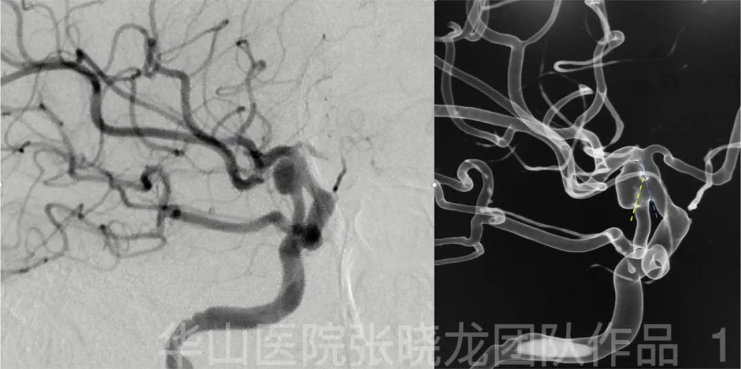
Figure 1. The aneurysm protruded laterally as the Pcom artery heading medially. Therefore two microcatheters were shaped with opposite spiral curve. 动脉瘤向外侧突出,后交通动脉朝向内侧。因此,两根微导管塑形成相反的螺旋形
01
Operation
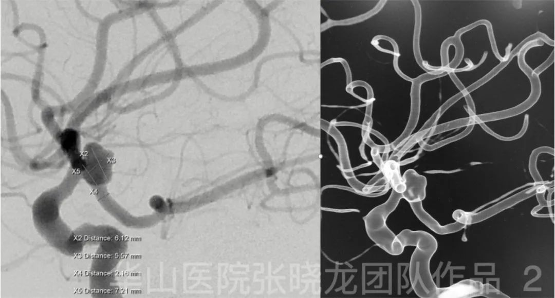
Figure 2. Measurement. 测量
Video 3. After slightly withdrawing the microcatheter, the Transend 205 microwire was advanced into the Pcom artery. Then the microwire tip was re-shaped into an acute curve and was advanced further to PCA. The stenting microcatheter was advanced to distal curve to deploy the stent stably. 微导管稍回撤后,Transend 205微导丝进入后交通动脉;随后,微导丝尖端重新塑形成急弯,并进一步到达大脑后动脉内。支架微导管向远端弯曲段推送,从而使支架稳定释放。
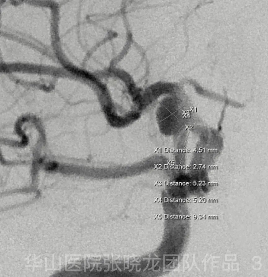
Figure 3. Measurement on the working projection of embolization. 栓塞工作角度上的测量。
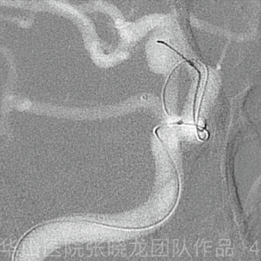
Figure 4 GIF. Echelon-10 for coiling was navigated to the aneurysm sac with the guide of 0.014 microwire. The aneurysmal neck was blocked by the first microcatheter. 在0.014微导丝引导下,将Echelon-10栓塞微导管置入动脉瘤腔内。通过第一根微导管填塞动脉瘤颈。
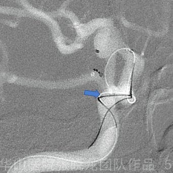
Figure 5 GIF. Solitaire 4mm*20mm was placed in the parent artery. The microcatheter for stenting was not retrieved. 将Solitaire 4mm*20mm支架置于载瘤动脉内。支架微导管不撤回。
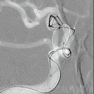
Figure 6 GIF. MicroPlex-10 7mm*30cm was inserted for framing. 填入MicroPlex-10 7mm*30cm弹簧圈成篮。
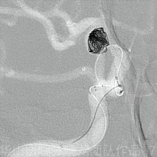
Figure 7 GIF. Electric detachment of Solitaire was failed. The stent was detached mechanically. General heparinization. Solitaire支架电解脱失败。支架机械解脱。行全身肝素化。
Figure 8 GIF. Angiography shows parent artery patent. 血管造影可见载瘤动脉通畅。

Figure 9 GIF. MicroPlex-10 5mm*15cm was inserted. But a longer coil may pack the dome better. 填入MicroPlex-10 5mm*15cm弹簧圈。但是一枚更长的弹簧圈或许能更好地栓塞动脉瘤顶部。
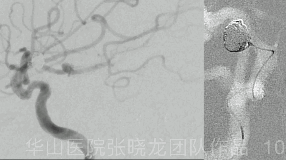
Figure 10 GIF. Angiography after inserting a MicroPlex-10 4mm*10cm coil shows partially protrusion of coil loops. 填入MicroPlex-10 4mm*10cm弹簧圈后,血管造影显示弹簧圈袢环部分脱出。
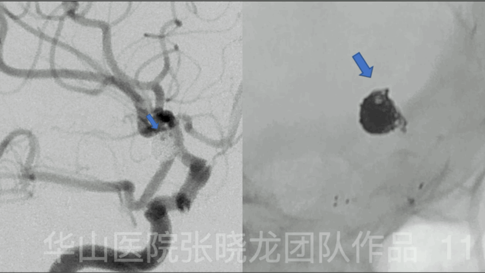
Figure 11 GIF. Angiography also indicates loose packing of superior part of the aneurysm. 血管造影也提示动脉瘤上部疏松栓塞。
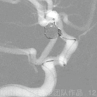
Figure 12 GIF. HyperSoft 1mm*4cm could not be inserted to the superior loose part of the aneurysm. The aneurysm neck was overpacked instead. HyperSoft 1mm*4cm弹簧圈不能填入动脉瘤上方疏松部分。而动脉瘤颈则过度栓塞。
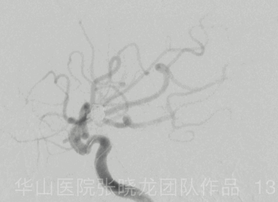
Figure 13 GIF. Rotation angiography shows overpacked aneurysm neck with loose packing of superior part of the aneurysm. The parent artery is patent. Tirofiban 13ml was administrated. 旋转血管造影显示动脉瘤颈过度栓塞、动脉瘤上部疏松栓塞。载瘤动脉通畅。给予替罗非班13ml。
02
Post operative complication
One hour after operation, the patient suffered from slurred speech, left limb muscle strength level I.
术后1小时,患者出现言语不清,左下肢肌力l级。
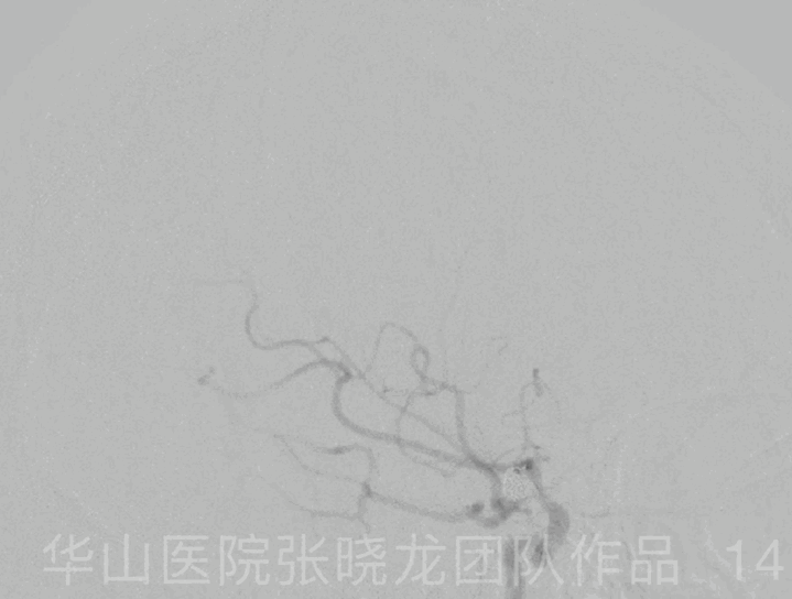
Figure 14 GIF. Emergency angiography shows intracranial vessels intact and parent artery patent. Thrombolysis of in-stent thrombosis was considered. Emergency Medication: General heparinization. Tirofiban 12ml iv. Tirofiban 20ml arterially during angiography. Nimodipine 2ml. 紧急血管造影颅内血管显示完整,载瘤动脉通畅。考虑支架内血栓溶解。紧急用药:全身素化。替罗非班12ml静注,替罗非班20ml造影时经动脉给药。并给予尼莫地平2毫升。
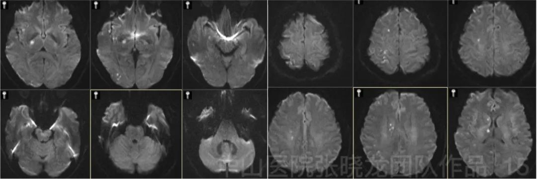
Figure 15. Upper limb muscle strength improved to III. Lower limb V-. MRI DWI shows scattered acute infarctions in the right hemisphere. 上肢肌力恢复至IlI级。下肢肌力V-级。MRI DWI显示右侧大脑半球散在急性梗塞灶。
03
Post operation
PE: GCS 15, left upper limb III-, left lower limb V-.
Medication: Aspirin 100mg qd, Clopidogrel 75mg qd for 3 months, Amlodipine 5mg qd.
查体:GCS 15,左上肢肌力IlI级,左下肢肌力V-级。
用药:阿司匹林100mg qd,氯吡格雷75mg gd 3个月,氨氯地平5mg qd。
04
Summary
Aneurysm size: 4.51mm*5.23mm; Neck: 2.94mm
Stent: Solitaire 4mm*20mm
Coils: Microplex-10 7mm*30cm, 5mm*15cm, 4mm*10cm (2), Hypersoft 1mm*4cm
For a primitive Pcom artery aneurysm, the neck should not be overpacked. Small thrombus formed in the initial Pcom artery could be flushed into MCA and ACA territory. The thromboembolic events could lead to a disaster.
After the operation, the patient suffered from left weakness, indicating thrombosis in the stent. General heparinization was performed.
动脉瘤尺寸:4.51mm*5.23mm;瘤颈:2.94mm
支架:Solitaire 4mm*20mm
弹簧圈:Microplex-10 7mm*30cm, 5mm*15cm, 4mm*10cm (2), Hypersoft 1mm*4cm
由于动脉瘤起源于原始后交通动脉,计划将Solitaire支架置于后交通动脉。不需要使用Y型支架。
对于原始后交通动脉瘤,瘤颈不应过度栓塞。因最初在后交通动脉形成的小血栓可进入大脑中动脉和大脑前动脉供血区域,血栓栓塞事件可导致灾难性的后果。
术后患者左侧肢体无力,提示支架内血栓形成,行全身肝素化。
05
Follow up records
Long term follow up (2 year): sudden onset of left limbs weakness and dizziness (without numbness) 2 weeks ago. PE: left limbs muscle strength V. Medication: Amlodipine 5mg qd, Zopiclone1/2-1/3# qn.
短期随访(3个月):查体:左侧一过性肢体麻木,左侧肢体肌力V级。用药:氨氯地平5mg gd,阿司匹林100mg qd。
长期随访(2年):2周前突发一过性左侧肢体无力、头晕(无肢体麻木)。查体:左侧肢体肌力V级。用药:氨氯地平5mg gd,佐匹克隆1/2-1/3# qn。
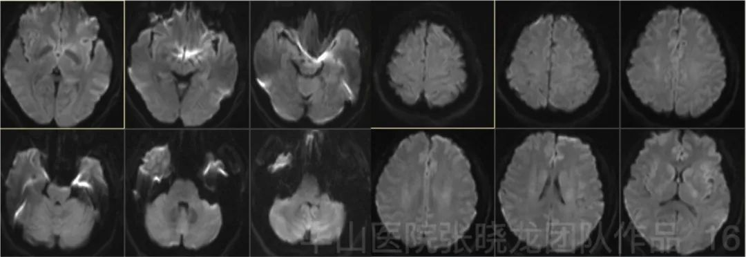
Figure 16. Follow up MRI DWI (3 months) shows no acute infarctions. 3个月随访MRI DWI未见急性梗塞灶。
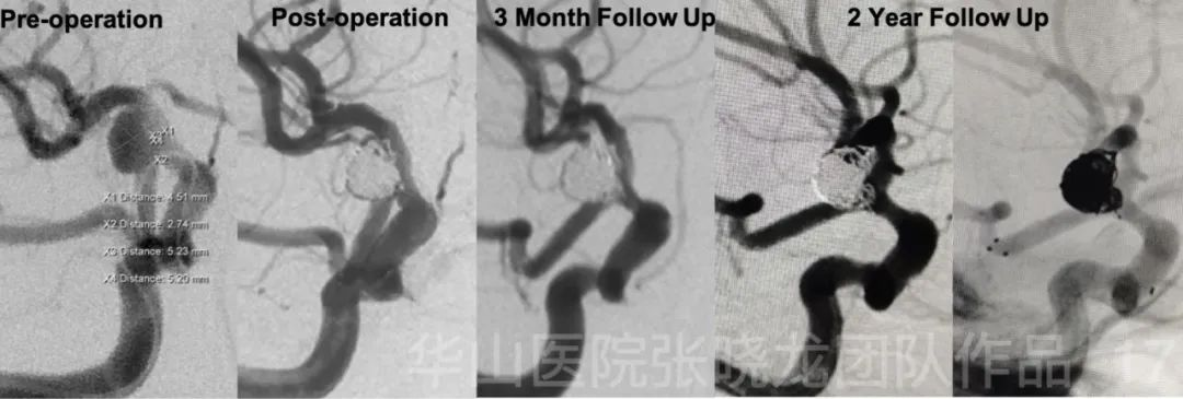
Figure 17. No relapse of the aneurysm during follow up. 随访期间未见动脉瘤复发。
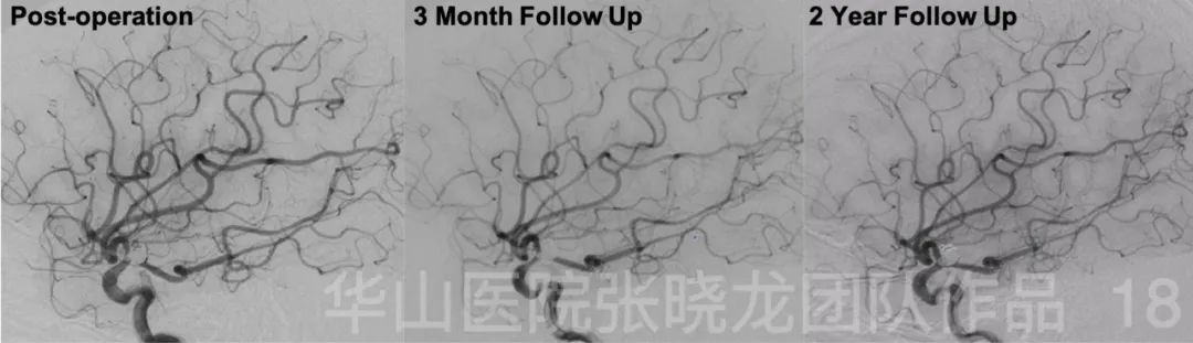
Figure 18. Follow up angiographies show the patent of the parent artery and the intact of intracranial vessels. 随访血管造影显示载瘤动脉通畅,颅内血管完整。
06
Summary
![]()
