Basilar artery plaque contained fat issue and pontine infarction
基本信息 Basic Information
男,67岁
67-year-old male
症状:突发右侧肢体无力
Symptoms: sudden weakness of the right limb
高血压、糖尿病和高胆固醇血症
Hypertension, diabetes and hypercholesterolemia
影像资料
Imaging Data
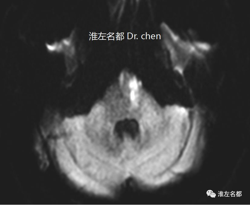
△DWI:左侧脑桥梗死
△DWI:left pontine infarction
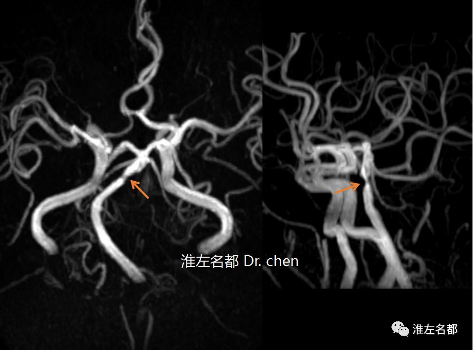
△头颅3D-TOF-MRA:基底动脉狭窄(橙箭)
△Cranial 3D-TOF-MRA: basilar artery stenosis (orange arrow)
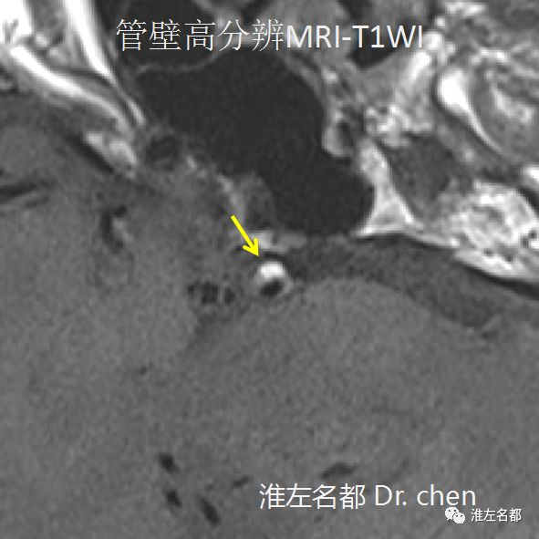
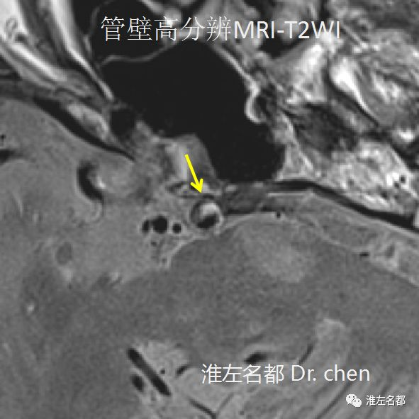
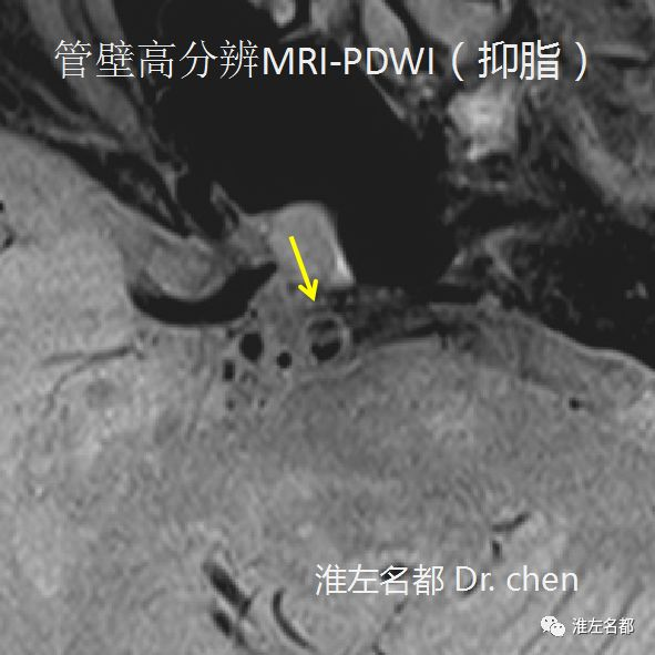
△管壁高分辨MRI:基底动脉局部管壁偏心增厚(黄箭),其内信号不均匀(提示斑块);PDWI(质子加权成像)抑脂序列可见临近管腔的条样等信号纤维冒;T1WI和T2WI于增厚管壁局部检测到小片状高信号,此高信号区域在PDWI抑脂序列呈低信号,提示脂肪组织
△ High resolution vessel wall MRI: an eccentric thickening of the vessel wall was detected in the basilar artery (yellow arrow), it had heterogeneous signal (atherosclerotic plaque); the proton density weighted image (PDWI) with fat supspanssion showed a strip-like plaque fiber cap with iso-signal intensity; a partial area of this plaque spansented with a hyper-signal intensity on T1WI andT2WI, but with a hypo-signal intensity on PDWI with fat supspanssion, which prompted that this area had the fat tissue.