


去年4月写了猴年第一贴《创刊号——大脑表面解剖与深部投影及其应用一例》,7月在《侧裂——解剖小汇总》里涉及了一些脑深部定位的知识,但真的是纸上谈来终觉浅。今天在猴年的小年夜顺顺利利夜出,决定再次以颅脑解剖定位这个问题来写个“拜年贴“”。在影像导航技术越来越发达的现在,我始终信奉Rhoton教授“神经外科医师必须有一双see-through-X-ray的眼睛”的观点;没有这双眼睛,面对再先进的术前影像也无法准确读片;没有这双眼睛,摆不好体位导航也只是空摆设。平时工作中,用到的解剖定位方法主要是基于Rhoton教授的那副经典简图(下图),中央沟、侧裂可以大致定位,但是对于更细化的结构可能就不太够用了。因此才有了这篇进阶版,内容完全是基于来自巴西圣保罗大学的颅脑解剖大师Ribas教授的经典文献《Surgical anatomy of microneurosurgical sulcal key points》和一个意大利解剖网站http://3dneuroanatomy.com的在线文献《craniometric points of the skull and the cerebral cortical surface》进行归纳总结。引用的图片均为红蓝3D格式。希望来年能在这方面继续进步。
1
十大定位点概览
大脑皮层的沟回复杂且存在变异,但有那么几条脑沟或几个点相对固定,它们往往是蛛网膜池在皮层表面的局部扩大处,并且存在较重要的深部结构对应关系。同样的,颅骨表面也存在数个明显且恒定的骨性结构。奇妙之处在于,这些颅骨(cranial)结构和皮层(cortical)结构竟然存在着许多一一对应的解剖关系,似乎是造物主在冥冥之中就为人类提供了便捷的外科学定位依据。
1.1 颅骨定位标志
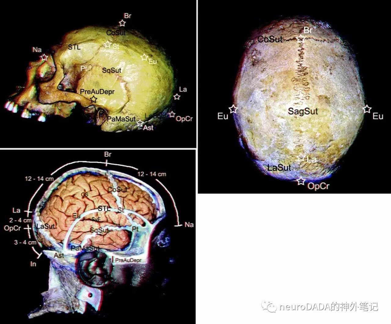
Ast, asterion,星点
Br, bregma,前囟
CoSut, coronal suture,冠状缝
Eu, euryon,颅阔点(顶结节)
FZgS, frontozygomatic suture,额颧缝
In, inion,枕外粗隆
La, ; LaSut, lambdoid suture,人字缝
Na, nasion,鼻根点
OpCr, opisthocranion,枕后点
PaMaSut, parietomastoid suture,顶乳缝
PreAuDepr, preauricular depression,耳前压迹
Pt, pterion,翼点
SagSut, sagittal suture,矢状缝
SqSut, squamous suture,鳞状缝
St, Stephanion,冠状点
STL, superior temporal line,颞上线
1.2皮层定位标志
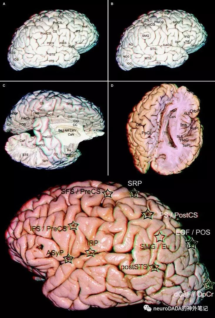
ASyP, anterior sylvian point,前侧裂点
dCaF/OpCr, distal calcarine fissure point / opisthocranion,距状裂远端(枕后点深面)
EOF/POS, external occipital fissure medial point / parieto-occipital sulcus,枕外裂内侧点=顶枕沟在脑内侧面的最上端
IFS/PreCS, inferior frontal sulcus and precentral sulcus meeting point,额下沟与中央前沟交点
IPS/PostCS, intraparietal sulcus and postcentral sulcus transitional or meeting point,顶内沟与中央后沟交点
IRP, inferior Rolandic point,下中央沟点
postSTS, superior temporal sulcus posterior segment and extremity,颞上沟后端
SFS/PreCS, superior frontal sulcus and precentral sulcus meeting point,额上沟与中央前沟交点
SMG/EU, supramarginal gyrus / Euryon,缘上回(颅阔点深面)
SRP, superior Rolandic point,上中央沟点
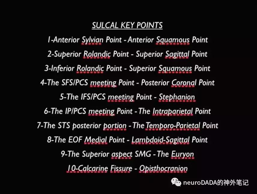
1.3十组颅-皮层定位点对应关系
2
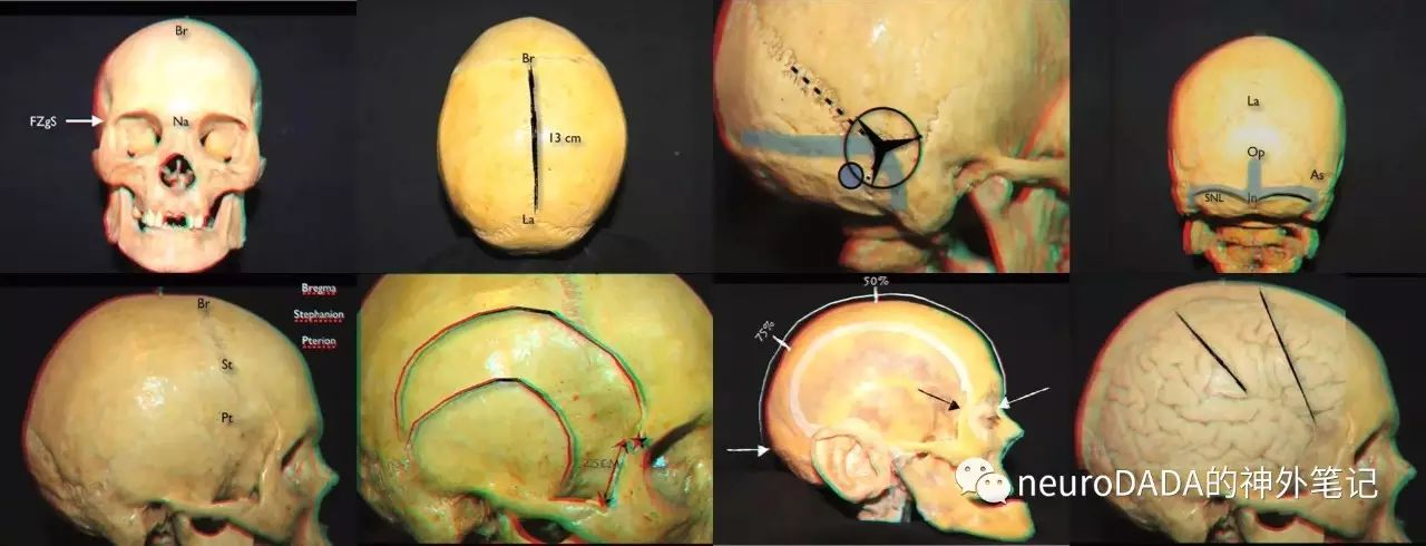
额颞开颅相关定位点(3对)
前侧裂点 ASyP——前鳞状点 ASqP
下中央沟点 IRP——上鳞状点 SSqP
额下沟与中央前沟交点 IFS/PreCS——冠状点 St

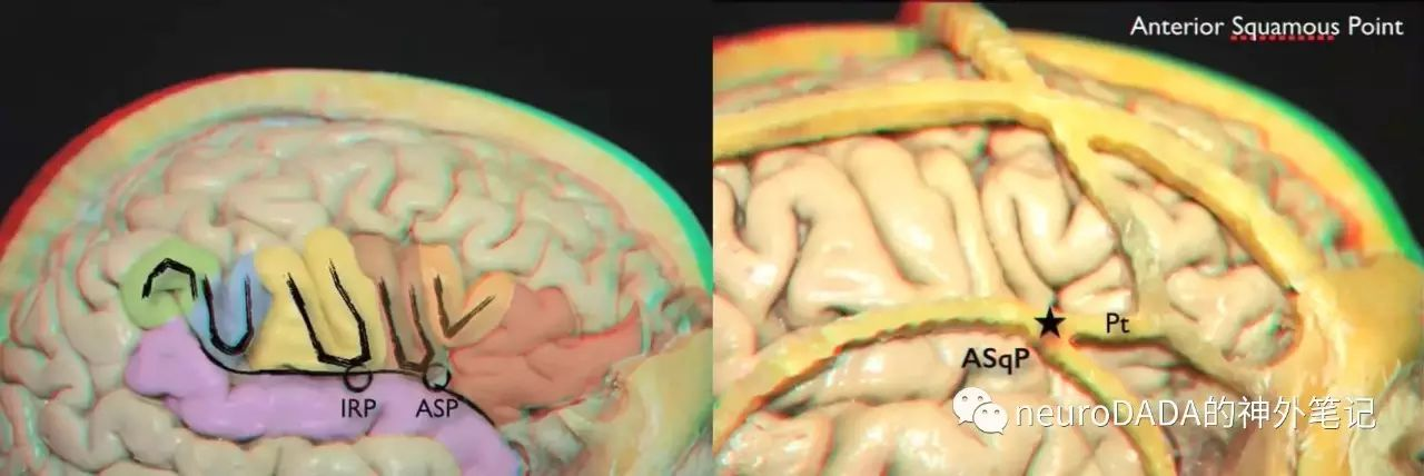
2.1前侧裂点 ASyP——前鳞状点 ASqP
前侧裂点 anterior Sylvian point
额下回三角部回缩,其下方的侧裂池局部扩大
前鳞状点 anterior squamous point
翼点区域H形骨缝,鳞状缝前部与顶-蝶缝交界处
对应度
垂直面上70%,水平面上56%
临床意义
即Yasargil所谓的Sylvian point(侧裂点),解剖侧裂的最佳起始点
额叶盖部的三角区和盖区为Broca区
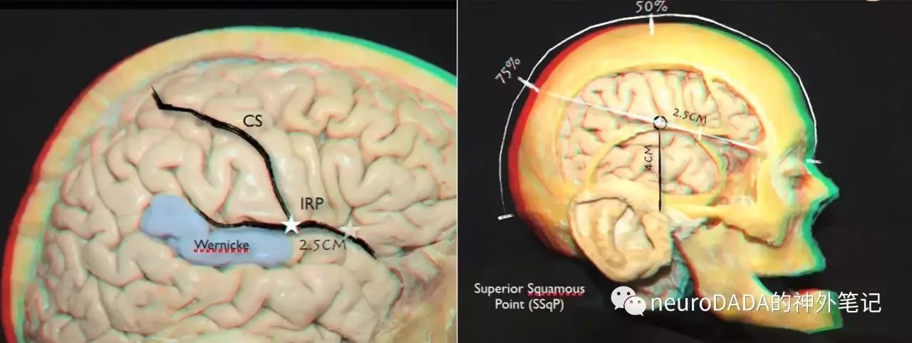
2.2下中央沟点 IRP——上鳞状点 SSqP
下中央沟点 inferior Rolandic point
中央沟(或其延长线)与侧裂的交点
沿侧裂,位于ASP后方2.5(2-3)cm
上鳞状点 superior squamous point
鳞状缝的最高点
鳞状缝与耳前压迹所在垂直线的交点,耳前压迹上方4(3.5-4.5)cm
耳前压迹 preauricular depression:颧弓上方、耳屏前方的凹陷
Rhoton定位法:侧裂线(额颧点、鼻根-枕外隆凸连线后三分之一点连线)上,翼点后方2.5cm
对应度
65%位于侧裂内,29%恰位于IRP
临床意义
对应侧裂下方的颞横回的Heschl s回(听觉中枢)前缘,与之相对的是中央后回
其后方的颞横回、颞上回和部分角回构成了Wernicke区
界定了颞叶上表面的极平面polar planum和颞平面temporal planum
对应中央沟下方的中央下回subcentral gyrus,又称inferior Broca’s frontoparietal plis de passage
2.3额下沟与中央前沟交点 IFS/PreCS——冠状点 St
额下沟与中央前沟交点 IFS/PreCS
额下沟平行于侧裂
额下沟后端可位于中央前沟前方、重叠、后方
冠状点 stephanion
冠状缝与颞上线的交点
沿冠状缝,距离前囟7.8(7-8.5)cm
对应度
70%冠状点位于额下沟
30%冠状点位于中央前沟,53%位于中央前沟前方
IFS/PreCs通常位于St后方<2cm距离内
临床意义
后方正对着中央前回的面部运动区
与ASP、IRP一起界定额下回的高度、Broca区的大致范围

3
额上部与中央区开颅相关定位点(2对)
额上沟与中央前沟交点 SFS/PreCS——冠状后点 PCoP
上中央沟点SRP——上矢状点SSaP
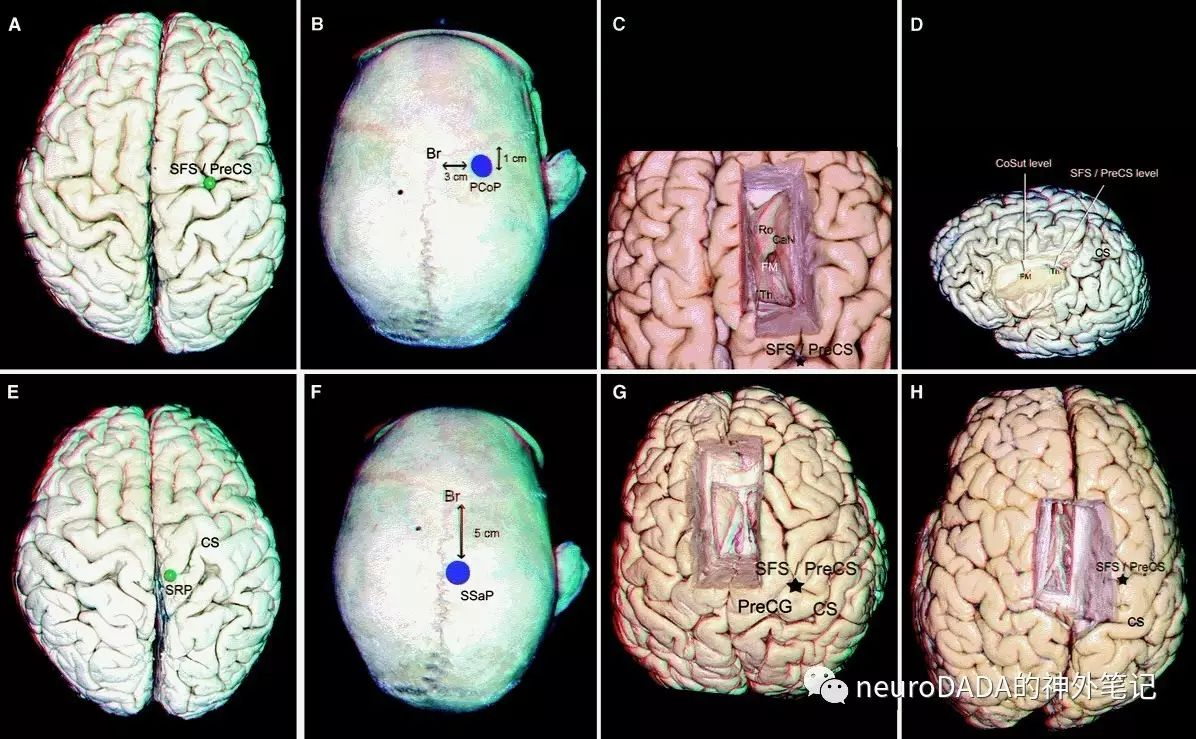
3.1额上沟与中央前沟交点 SFS/PreCS——冠状后点 PCoP
额上沟与中央前沟交点 SFS/PreCS
额上沟平行于纵裂
额上沟后端可位于中央前沟前方、重叠、后方
冠状后点 posterior coronal point
冠状缝后方1cm,中线旁开3cm
对应度
81%冠状后点位于额上沟
25%冠状后点位于中央前沟
冠状后点一般对应于SFS/PreCS实际交点的偏前方(保证安全性,见下)
临床意义
后方正对着中央前回的手部运动区
经额上沟入路,切开额上沟的最远后界
冠状位上,平丘脑、侧脑室体部、室间孔略后方(冠状缝对应室间孔)
半球间经胼胝体入路,额叶牵开范围、胼胝体切开范围的最远后界,因此骨窗大部分位于冠状缝前

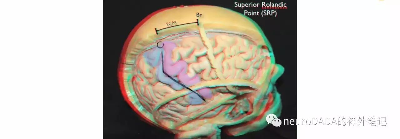
3.2上中央沟点SRP——上矢状点SSaP
上中央沟点 superior Rolandic point
上端总是与纵裂相交
上矢状点 superior sagittal point
前囟后方5cm
鼻根-枕外粗隆连线中点后方2cm
对应度
37.5%,上矢状点一般对应于SRP的偏前方
临床意义
半球间经胼胝体入路显露侧脑室体部,若以该点为中心则会偏后,而将显露胼胝体压部下方、穹窿联合后方的松果体区
4
顶叶入路相关定位点(3对)
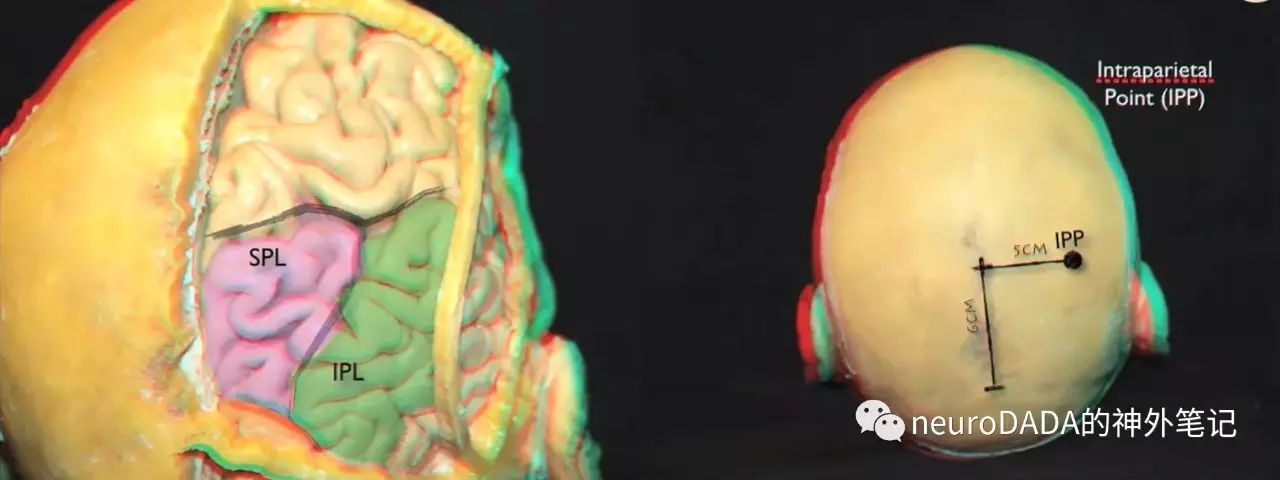
顶内沟与中央后沟交点 IPS/PostCS——顶内点 IPP
枕外裂内侧点或顶枕沟上端 EOFm/POS——人字缝/冠状缝交点 La/Sa
缘上回上部 SMG——颅阔点(顶结节)Eu
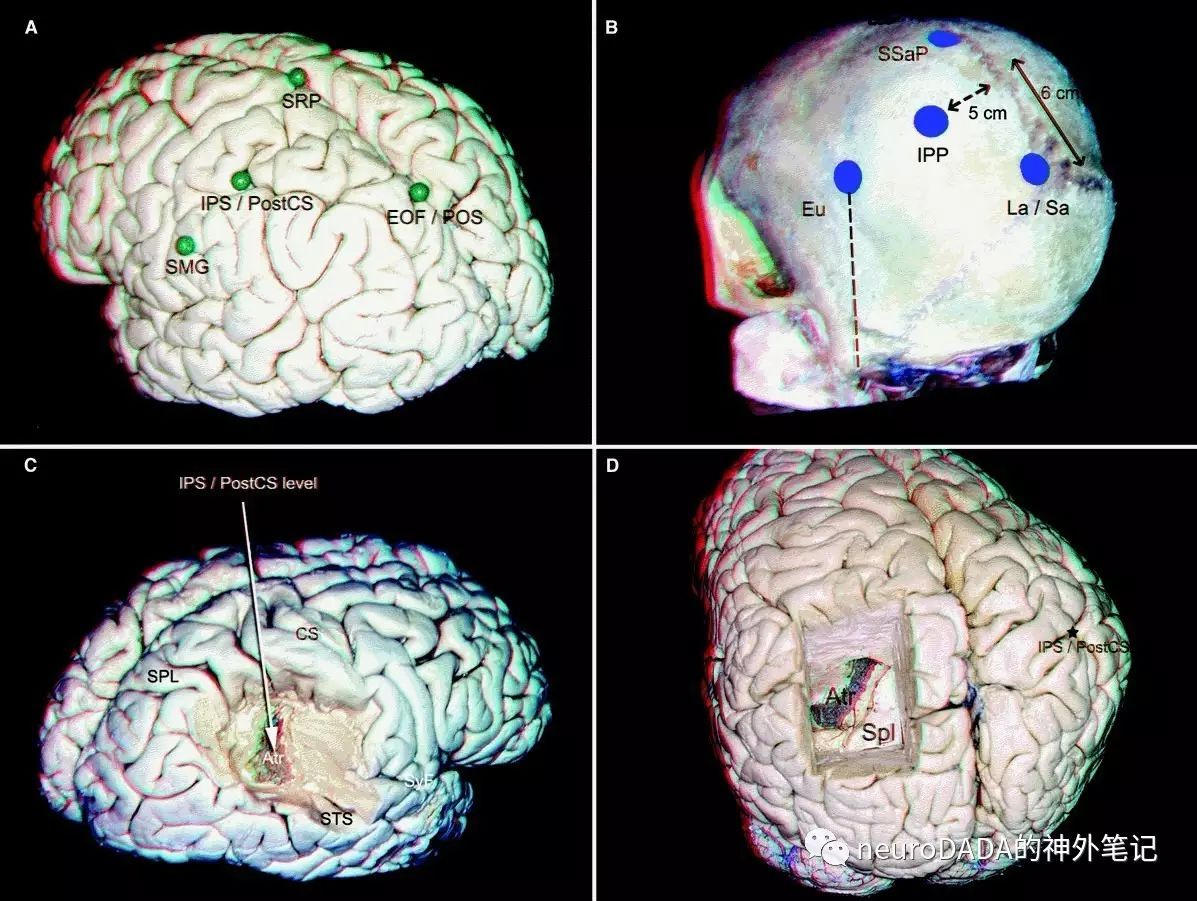
4.1顶内沟与中央后沟交点 IPS/PostCS——顶内点 IPP
顶内沟与中央后沟交点 IPS/PostCS
顶内沟大多平行于纵裂,也可垂直于纵裂
大多向前与中央后沟延续,也可孤立
将顶叶分为上方的顶上小叶和下方的顶下小叶,后者包含缘上回和角回
向后延续为分割枕叶的枕内回intraoccipital sulcus=枕横回transverse occipitalsulcus=枕上回superior occipital sulcus(具体见文末附)
顶内点 intraparietal point
人字缝/矢状缝交点前方6cm、中线旁开5cm
对应度
已较初始的顶内点(前方5cm、旁开4cm)对应度高
临床意义
经顶内沟入路,切开脑沟的最远前界
冠状位上,该点位于侧脑室房部后方,基本平胼胝体压部,少数位于其后方,因此向前30°角可进入侧脑室房部,少数需45°
顶内沟和中央后沟富含皮层静脉
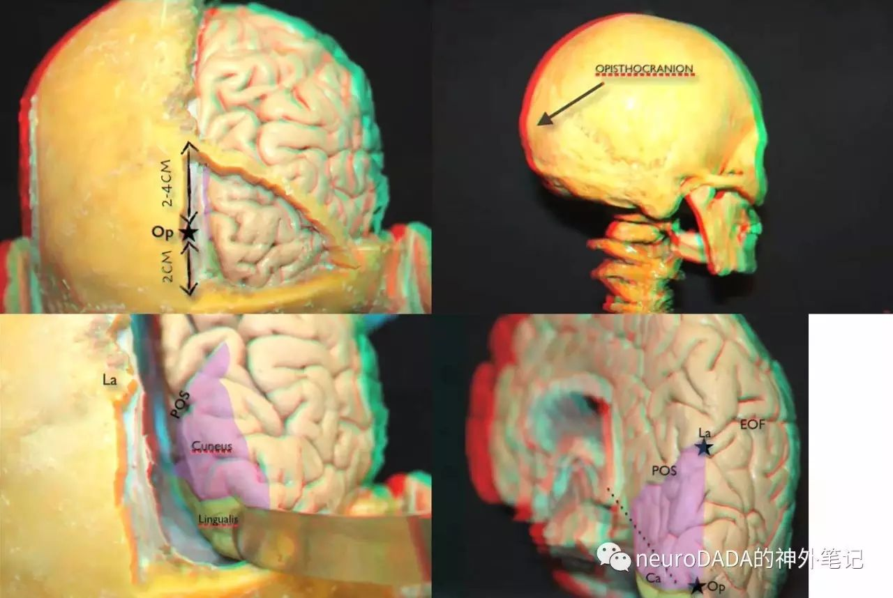
4.2枕外裂内侧点或顶枕沟上端 EOFm/POS——人字缝/冠状缝交点 La/Sa
枕外裂内侧点或顶枕沟上端 EOFm/POS
枕外裂external occipital fissure长度约2.2cm
人字缝/冠状缝交点 lambdoid/sagittal point
位于枕后点上方3(2-4)cm,位于前囟后方13(12-14)cm,位于鼻根后方25(24-26)cm
对应度
50%对应,44%骨性标志稍靠后
临床意义
顶枕叶的分界点
距离中央后沟约4cm,即楔前叶(顶叶内侧面)的纵向长度
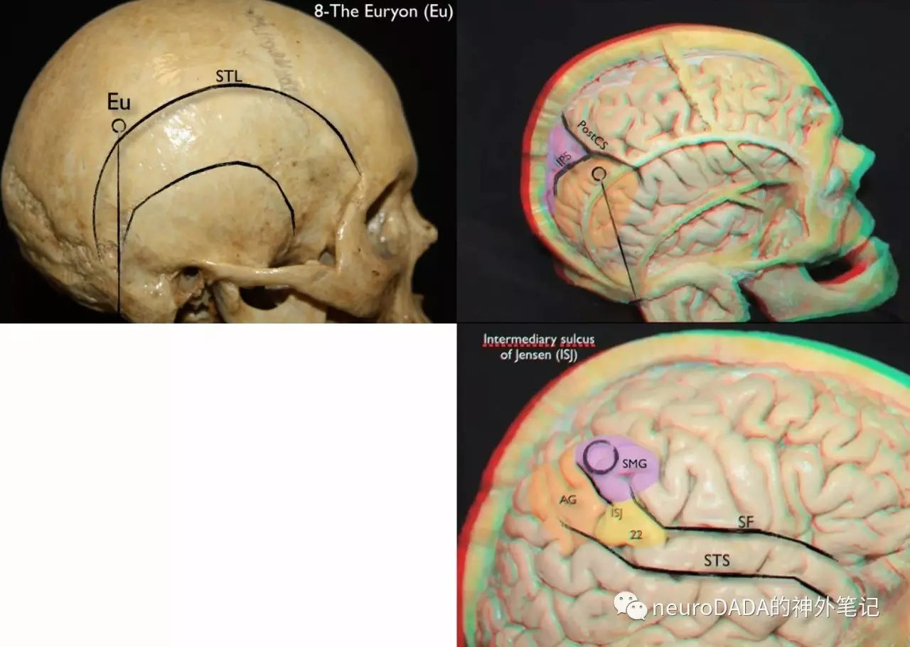
4.3缘上回上部 SMG——顶结节(颅阔点)Eu
这部分是单从颅骨标记点开始探讨的
颅阔点Euryon
顶结节parietal tuberosity最为突出的点
大多数位于颞上线上方
大多数位于一条垂直线上,该垂直线始于乳突尖,经过顶乳缝-鳞状缝移行点
位于IPP前下方
对应关系
其深面为缘上回上部,大多数位于上后部
位于后侧裂点posterior Sylvian point的上方和后方各约2.5(2-3)cm
位于中央后沟的后方约2(1.5-2.5)cm
位于Jensen中间沟 intermediary sulcusof Jensen (ISJ) 的前方约1.5cm,ISJ是缘上回和角回的分界线
临床意义
优势半球Eu下方为顶叶语言区(侧裂上方1-4cm,中央后沟后方2-4cm)
5
后颞部开颅相关定位点(1对)
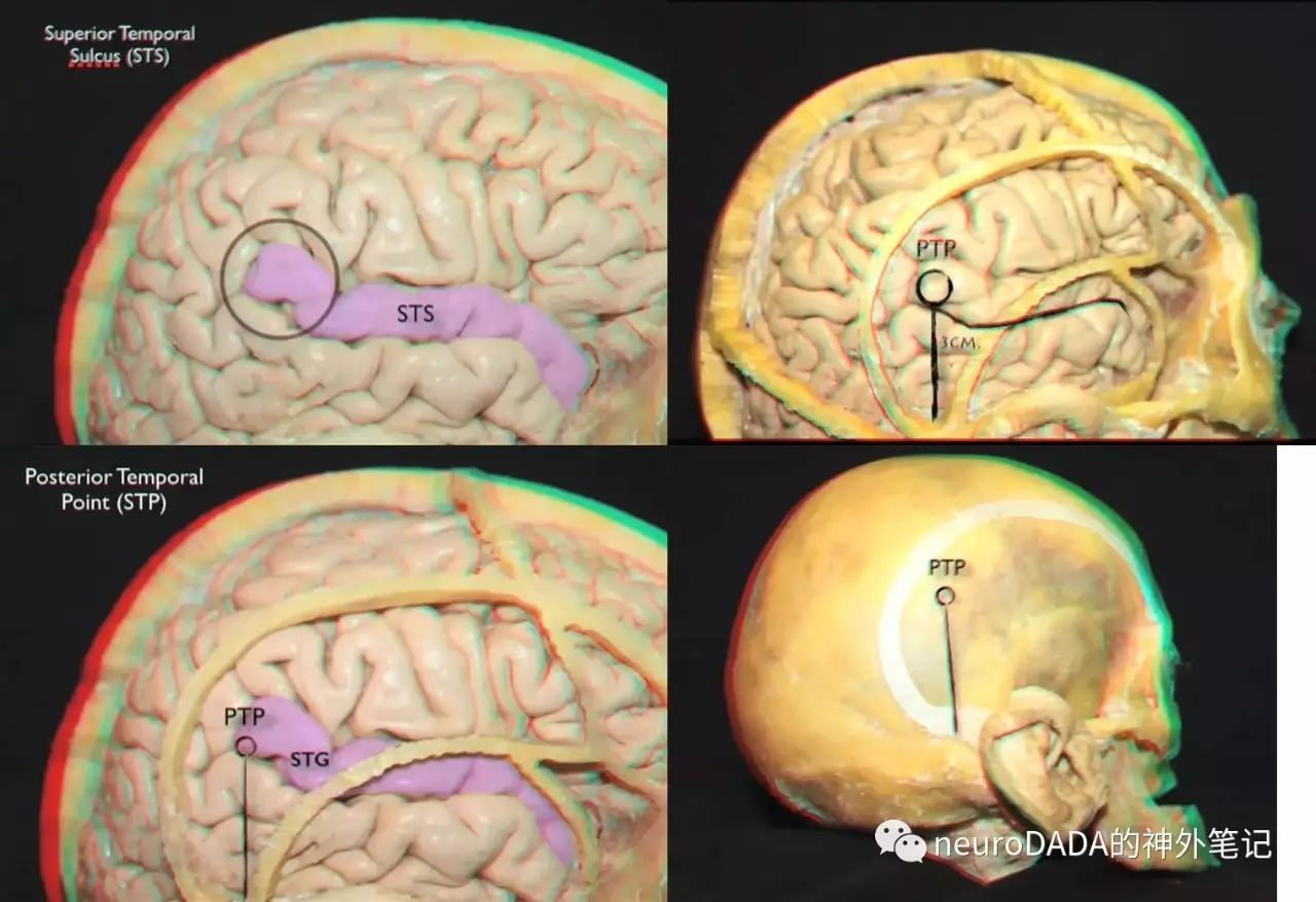
颞上沟后端 postSTS——颞顶点 TPP 或 颞后点 PTP
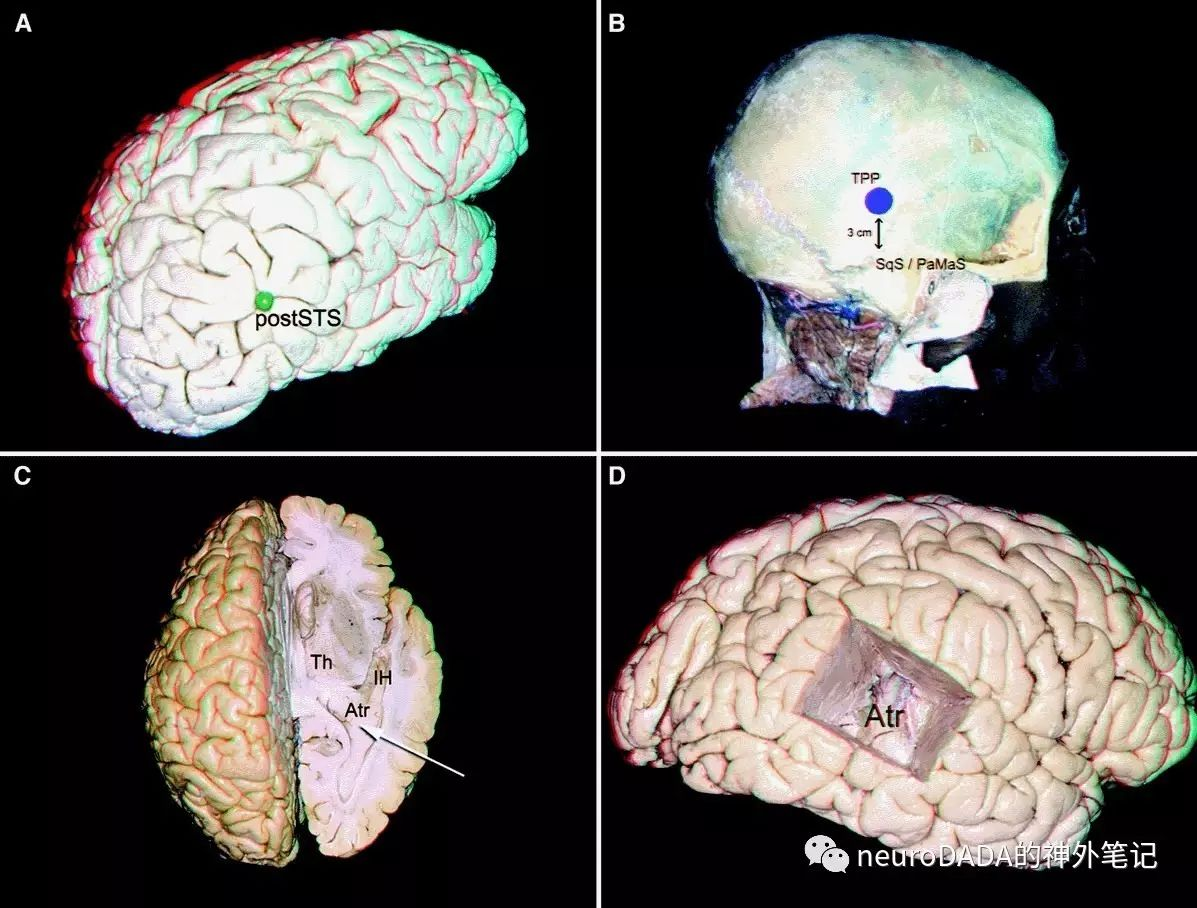
5.1颞上沟后端 postSTS——颞后点 PTP
颞上沟后端 posterior point of the posterior segment of the superior temporal sulcus
大多与颞上沟前部延续,少数不延续或独立
位于后侧裂点的后下方各约2-3cm
颞后点 posterior temporal point 或 颞顶点 temporoparietal point
位于顶乳缝-鳞状缝移行点的垂直上方3cm
位于颞上线后部下方
对应度
80%相对应
临床意义
经颞上沟可暴露整个侧脑室颞角
向前45°(少数30-40°)经postSTS可暴露侧脑室房部,尤其适合经脉络裂进入四叠体池的病变
可用于定位后侧裂点
易损伤语言传导束,对视觉传导束轻度影响
顶乳缝-鳞状缝移行点为颞骨嵴后缘、天幕附着点、乙状窦-横窦转角前缘,开颅需注意
6
枕部开颅相关定位点(1对)
距状裂后端 dCaF——枕后点 OpCr
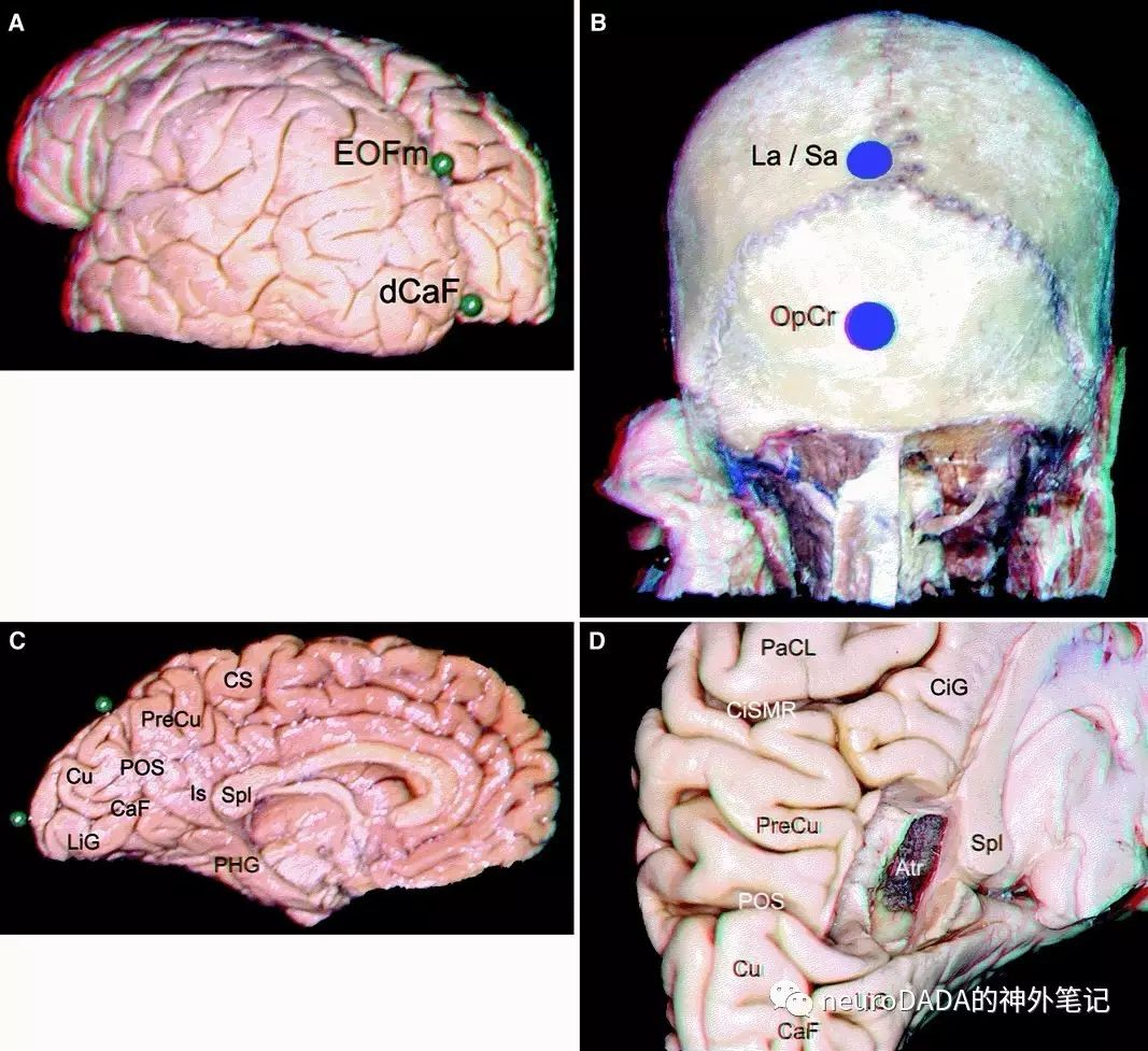
6.1距状裂后端 dCaF——枕后点 OpCr
距状裂后端 distal end of the calcarine fissure
距状裂在枕叶内侧面分隔上方的楔叶和下方的舌回
枕后点 opisthocranion
枕骨最突向后方的点
人字缝下方3(2-4)cm
枕叶底部上方2(1-3)cm,即舌回的纵向高度
枕骨粗隆上方3.5(3-4)cm
对应度
枕后点略高于距状裂后端上缘
临床意义
枕后点、距状裂后端、扣带回峡部、胼胝体压部基本位于一直线
Poppen入路骨瓣可以此为中心
注意距状裂上下的视皮层
附1、枕叶的界限
这是一个看似很简单但常常让人犯迷糊的问题,枕叶的界限必须从外侧面、内侧面、底面三个面上来说。
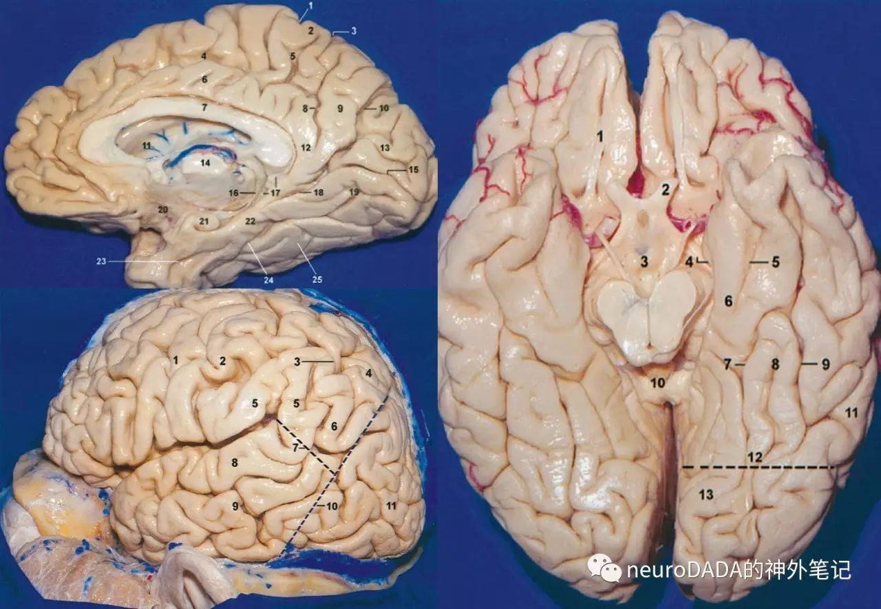
内侧面(左上图)
顶枕沟(parieto-occipital sulcus,10)分隔前方的顶叶(楔前叶 precuneus,9)和后方的枕叶(楔叶 cuneus,13 + 舌回 lingual gyrus,19)。距状沟(calcarine sulcus,15)分隔楔叶和舌回。舌回向前移行为颞叶内侧面的海马旁回(parahippocampal gyrus,22,右图6)。距状沟与顶枕沟的交点为颞叶内侧面的后界。海马旁回借侧副沟(collateral sulcus,24,右图7)及前方的鼻沟(rhinal sulcus,23,右图5)与梭状回(fusiform gyrus,又称枕颞回 occipitotemporal gyrus,25,右图8)相分隔。梭状回借枕颞沟(occipitotemporal sulcus,右图9)与颞下回(inferior temporal gyrus,右图11)分隔。梭状回向后续于枕下回(见附2)。
底面(右图)
底顶颞线(basal parietotemporal line,12)分隔后方的枕叶(舌回,13)和前方的颞叶底面。该线为一假想线,一端为顶枕沟下缘(与距状裂的交点),另一端为枕前切迹(preoccipital notch)。
外侧面(左下图)
外侧顶颞线(lateral parietotemporal line,10)分隔后方的枕叶和前方的顶叶、颞叶。该线为一假想线,一端为顶枕沟上端在半球外侧面切迹,即枕外裂(external occipital fissure)内侧端,另一端为枕前切迹。另外一条假想线为颞枕线(temporo-occipital line,7),分隔外侧面的顶叶和颞叶,一端为侧裂末端,另一端为外侧顶颞线的中点。
附2、枕叶的沟回
常规的解剖教科书上几乎没有涉及枕叶沟回的,对此各位解剖大师也历来存在争议。上文在述及顶内沟IPS时也提到了枕叶的沟回。这里引用同样来自Ribas教授的文献《The occipital lobe convexity sulci and gyri》,几幅简图大致看一下。
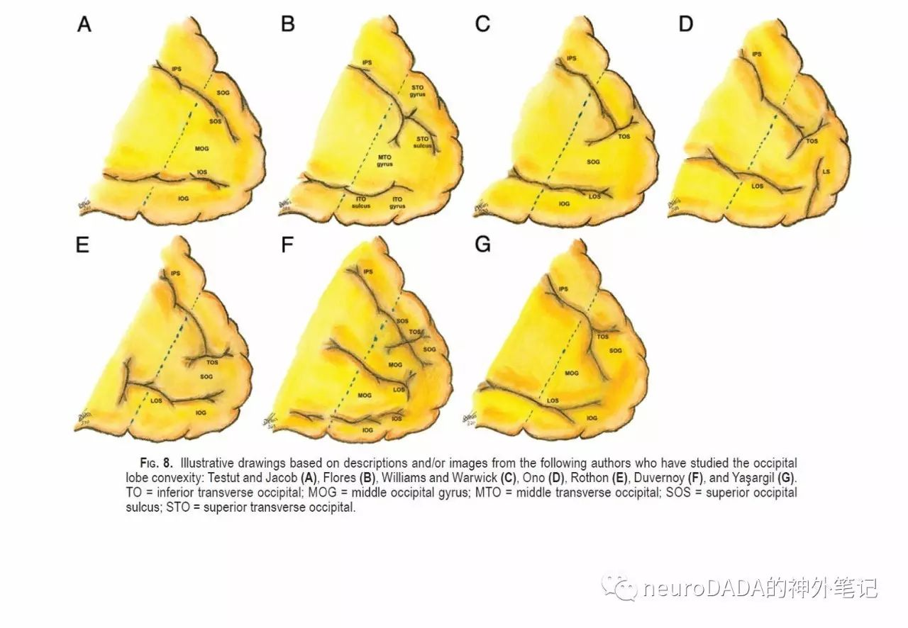
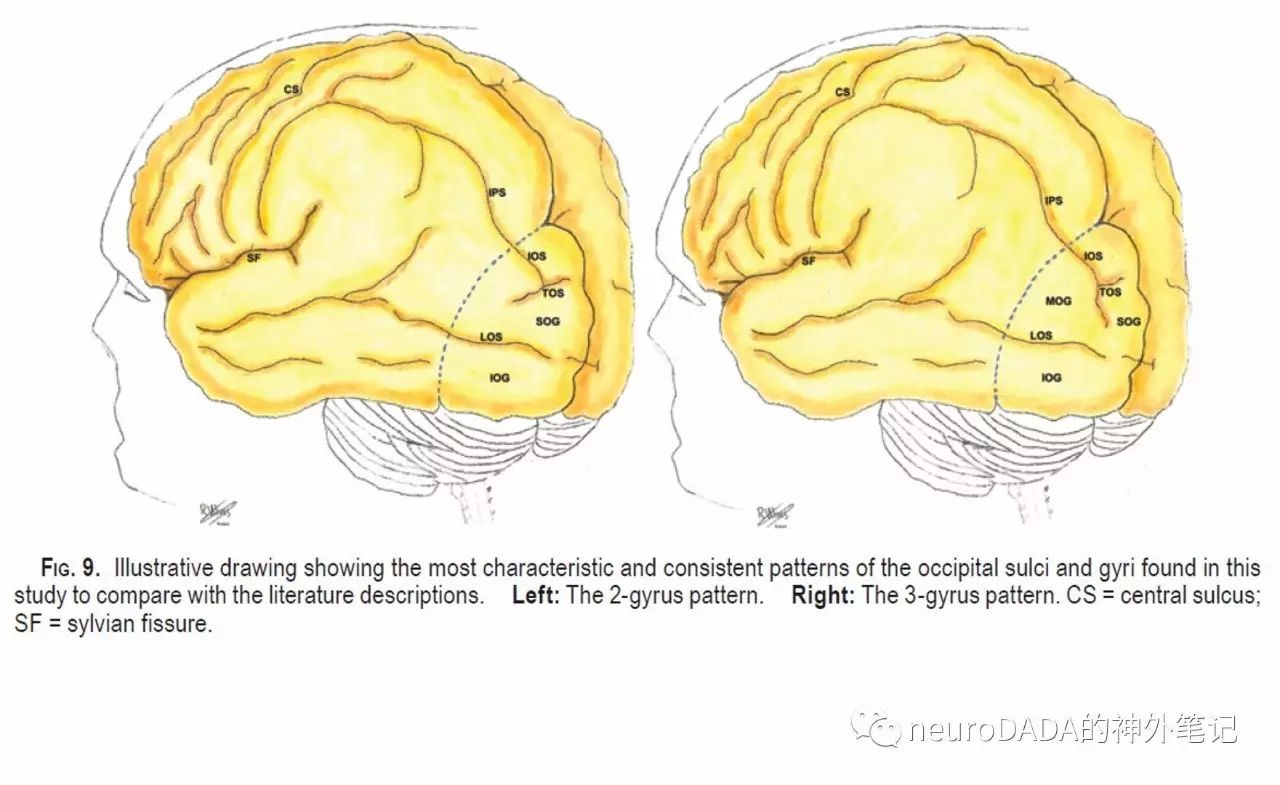
最后再引用一下Ribas教授的话:
To perform sophisticated cerebral microneurosurgical procedures, precise knowledge and proper identification of the brain sulci and gyri are mandatory in addition to fine microsurgical technique, and, obviously, neurosurgeons cannot be rely only on technological tools.
参考文献
1.Ribas GC, Yasuda A, Ribas EC, Nishikuni K, Rodrigues AJ, Jr. Surgical anatomy of microneurosurgical sulcal key points. Neurosurgery. 2006;59(4 Suppl 2):ONS177-210; discussion ONS-1.
2.Alves RV, Ribas GC, Parraga RG, de Oliveira E. The occipital lobe convexity sulci and gyri. Journal of neurosurgery. 2012;116(5):1014-23.
Wen HT, Rhoton AL, Jr., de Oliveira E, Cardoso AC, Tedeschi H, Baccanelli M, et al. Microsurgical anatomy of the temporal lobe: part 1: mesial temporal lobe anatomy and its vascular 3.relationships as applied to amygdalohippocampectomy. Neurosurgery. 1999;45(3):549-91; discussion 91-2.
4.craniometric points of the skull and the cerebral cortical surface, http://3dneuroanatomy.com
