


手术要点:
1.开颅前侧脑室外引流。
2.乳突后切口,延长至寰椎水平。
3.骨窗显露乙状窦,打开枕大孔外侧缘。
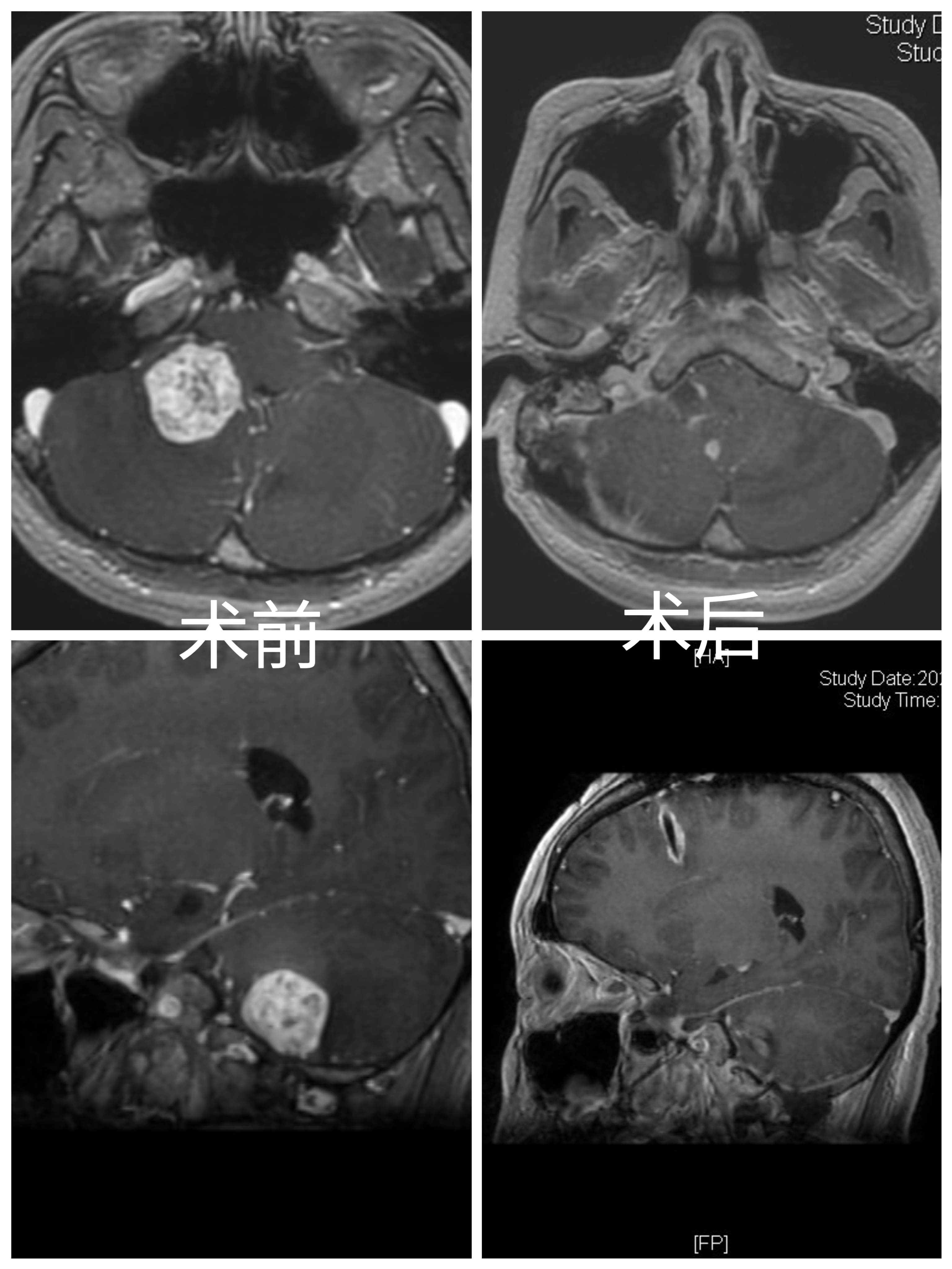
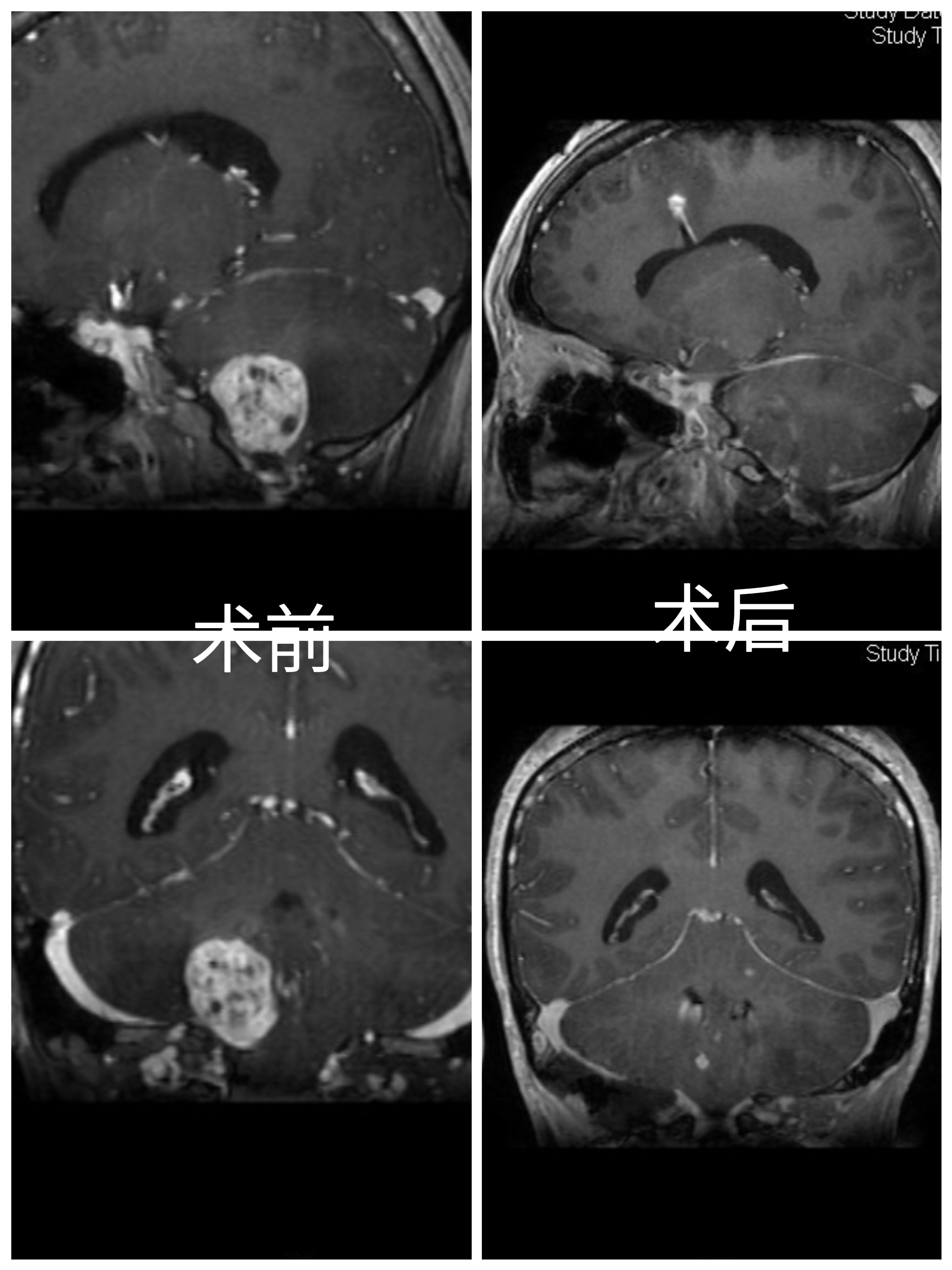
4.在小脑半球及小脑扁桃体外侧,切开脑组织,即见红色肿瘤。
5.沿肿瘤边缘分离,电凝切断血供,保护粗大引流静脉。
6.术中见肿瘤供血动脉,主要来源于小脑后下动脉的延髓外侧段、延髓近四脑室侧孔处动脉。
7.弱电凝延髓外侧细小血管,保护延髓正常血管。
8.临时阻断PICA,肿瘤血供明显减少。因肿瘤位于狭小空间,整体切除容易造成延髓损伤,而且不易电凝供血动脉。分块切除肿瘤,电凝切断延髓外侧段PICA,同时保护延髓及PICA。
9.严格沿边缘切除肿瘤,延髓表面静脉压迫止血。
10.术后病人轻度声音嘶哑、饮水呛咳。肢体活动正常。
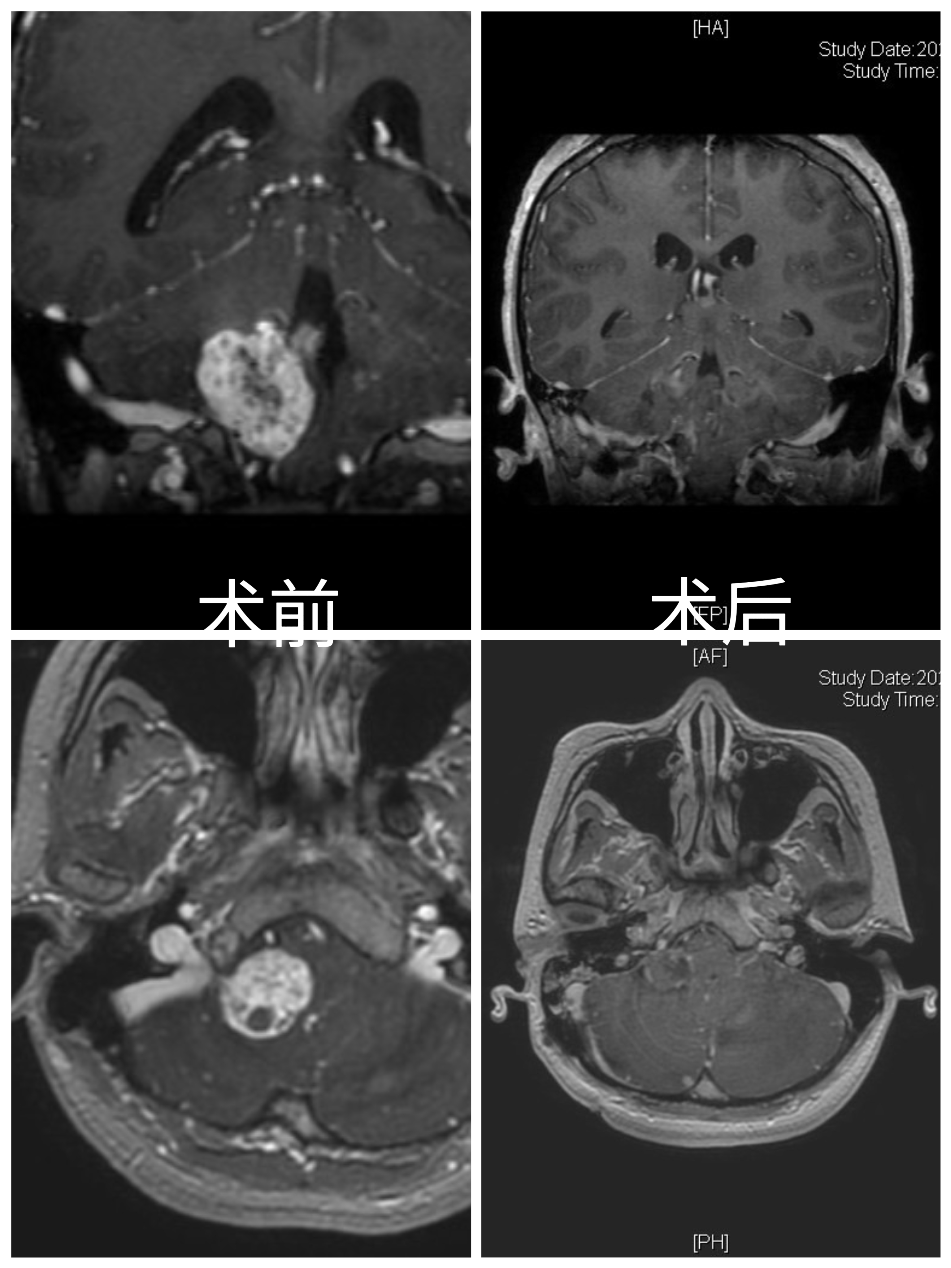
病例特点:
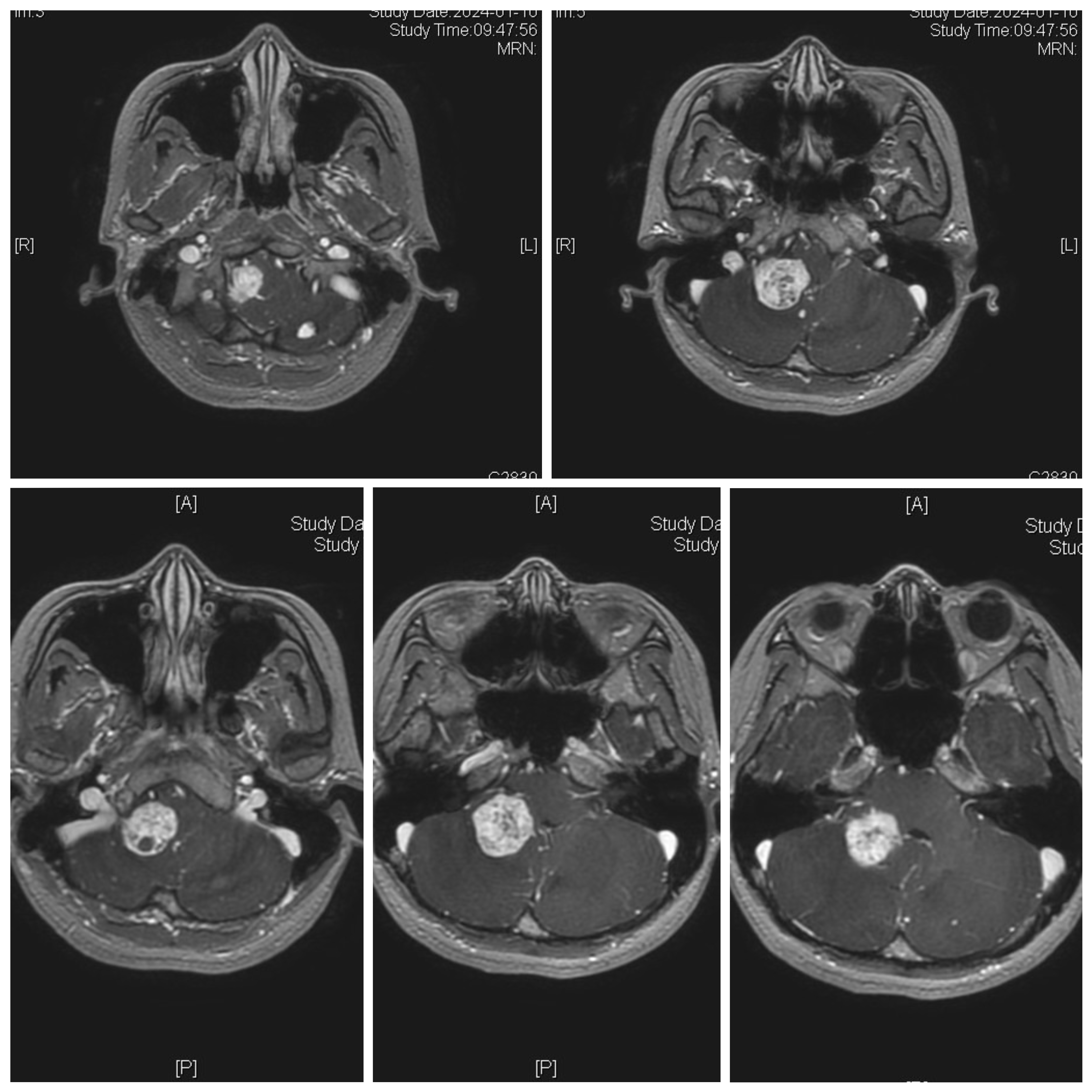
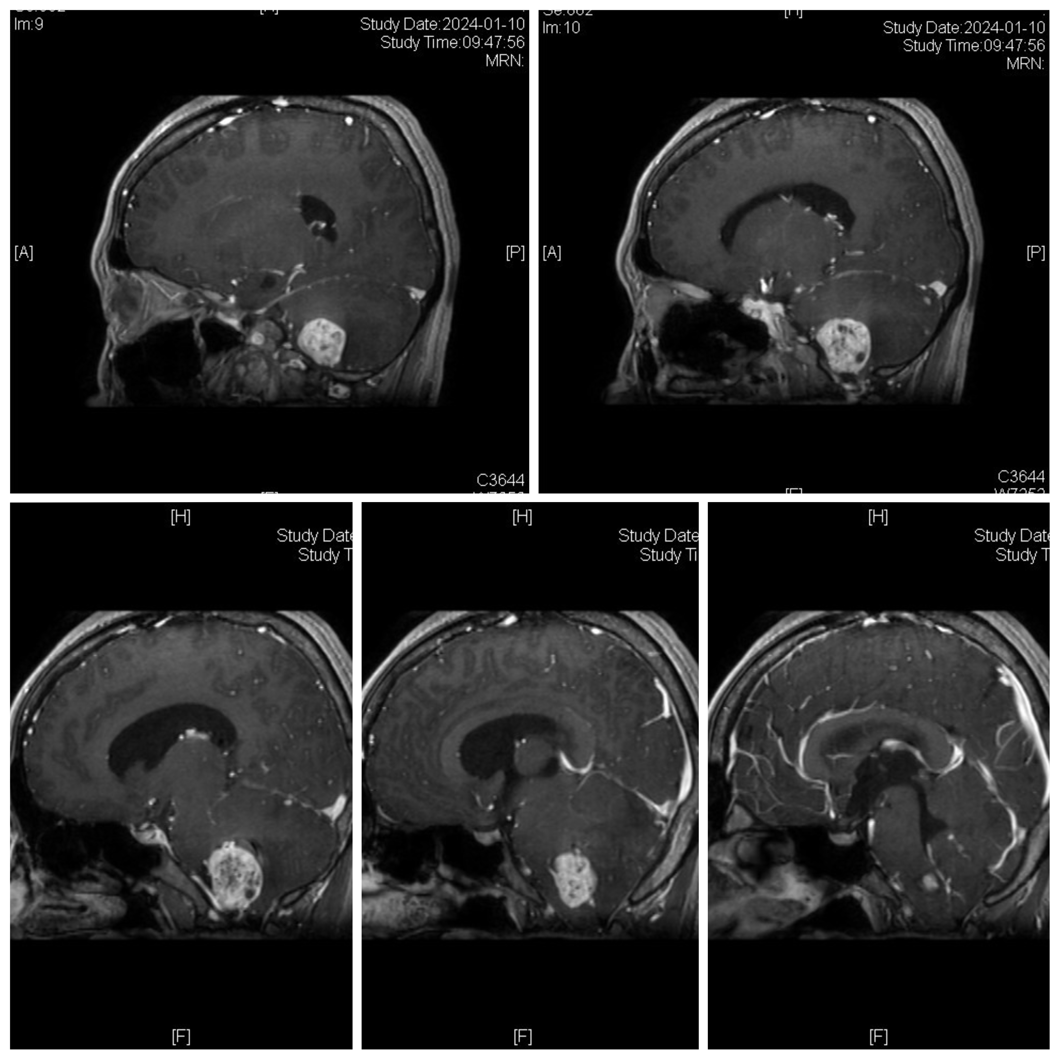
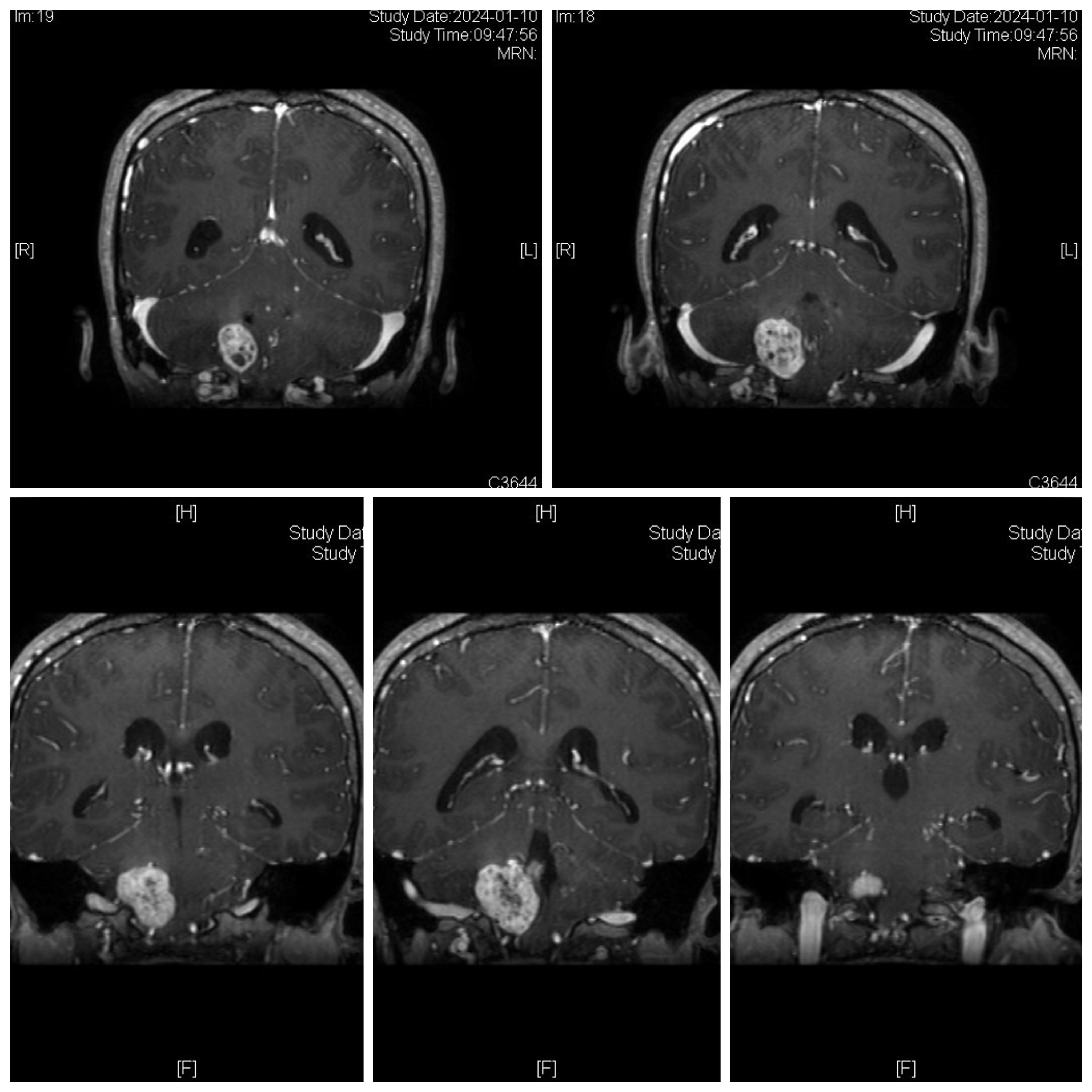
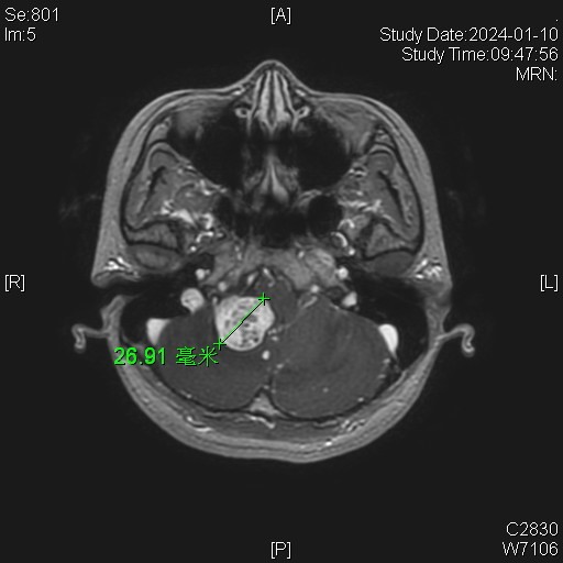
女性患者,26岁,因“间断性头晕1月余。”于2024-01-08入院。
患者1月余前感冒后出现头晕,伴头痛,伴恶心、呕吐。
家族史:患者母亲诊断多发小脑血管母细胞瘤,在我院手术治疗。
体检发现肝脏多发囊肿、右肾囊肿、胰腺多发囊肿。
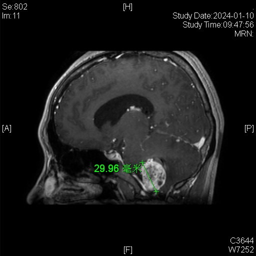
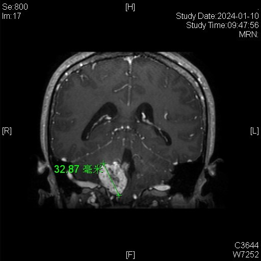
诊断:延髓血管母细胞瘤
von Hippel-Lindau综合征
Surgical points:
1. Preoperative external ventricular drainage on the lateral side of the brain.
2. mastoid incision, extended to the level of the atlas.
3. expose the sigmoid sinus through the bone window, and open the lateral edge of the foramen magnum.
4. On the lateral side of the cerebellar hemisphere and cerebellar tonsil, cut the brain tissue to reveal the tumor.
5. Separate along the edge of the tumor, coagulate and cut off the blood supply, and protect the large drainage veins.
6. During the operation, it was found that the feeding arteries of the tumor mainly originated from the lateral segment of the PICA ,near the foramen of the fourth ventricle of the medulla oblongata.
7. Weak coagulation of the small vessels on the lateral side of the medulla oblongata to protect the normal vessels of the medulla oblongata.
8. Temporarily block the PICA, and the blood supply of the tumor is significantly reduced. Due to the tumor located in a narrow space, it is difficult to remove the tumor as a whole ,and it is easy to damage the medulla oblongata. The tumor is removed in pieces, and the PICA on the lateral segment of the medulla oblongata is coagulated and cut off while protecting the medulla oblongata and PICA.
9. Strictly remove the tumor along the edge, and compress and stop bleeding on the surface of the medulla oblongata.
10. After the operation, the patient had mild hoarseness and drinking choking. The limb activity was normal.


视频1
视频2
